Review
. 2020 Jun 15;4(6):e20.00091.
doi: 10.5435/JAAOSGlobal-D-20-00091. eCollection 2020 Jun.

Metatarsal Pronation in Hallux Valgus Deformity: A Review

Affiliations
Review

Metatarsal Pronation in Hallux Valgus Deformity: A Review

Emilio Wagner et al. J Am Acad Orthop Surg Glob Res Rev. .

Erratum in

Abstract

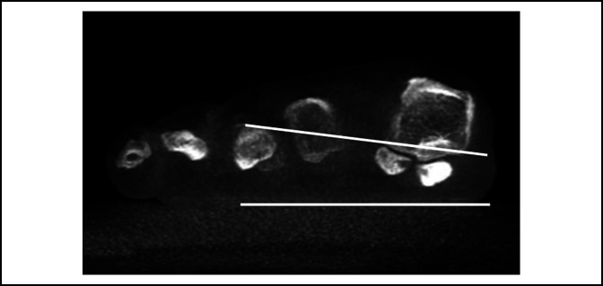
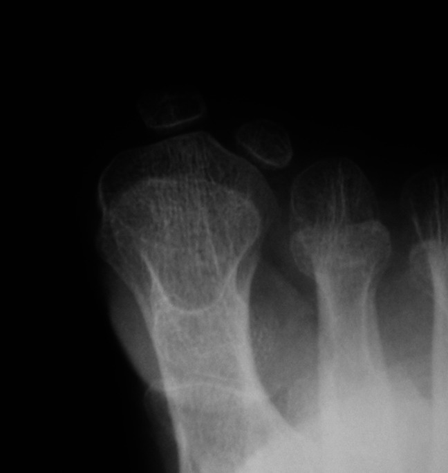
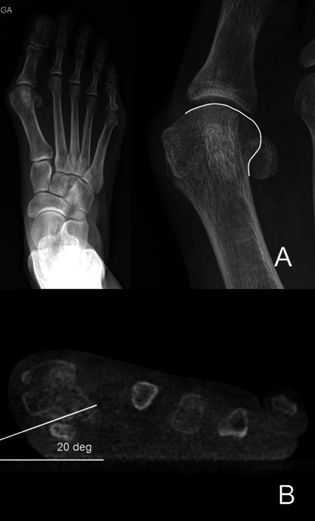
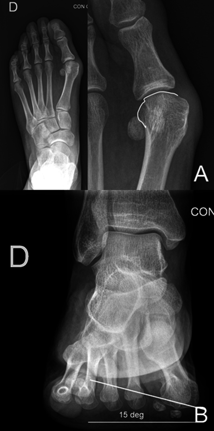
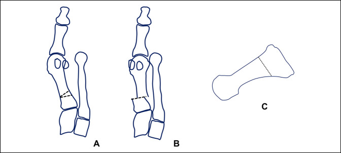
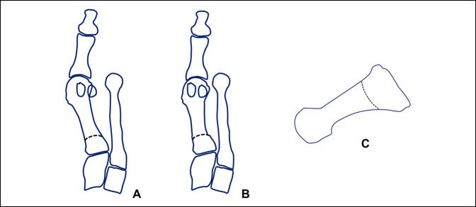
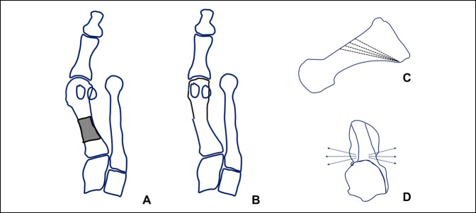
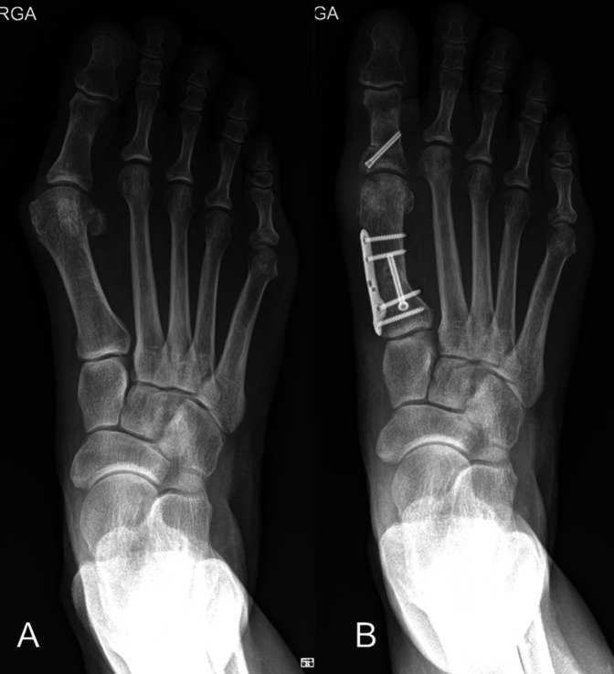
Hallux valgus deformity is a multiplanar deformity, where the rotational component has been recognized over the past 5 to 10 years and given considerable importance. Years ago, a rounded shape of the lateral edge of the first metatarsal head was identified as an important factor to detect after surgery because a less rounded metatarsal head was associated to less recurrence. More recently, pronation of the metatarsal bone was identified as the cause for the rounded appearance of the metatarsal head, and therefore, supination stress was found to be useful to achieve a better correction of the deformity. Using CT scans, up to 87% of hallux valgus cases have been shown to present with a pronated metatarsal bone, which highlights the multiplanar nature of the deformity. This pronation explained the perceived shape of the metatarsal bone and the malposition of the medial sesamoid bone in radiological studies, which has been associated as one of the most important factors for recurrence after treatment. Treatment options are discussed briefly, including metatarsal osteotomies and tarsometatarsal arthrodesis.


Conflict of interest statement

Neither of the following authors nor any immediate family member has received anything of value from or has stock or stock options held in a commercial company or institution related directly or indirectly to the subject of this article: Dr. E. Wagner and Dr. P. Wagner.

Figures

Figure 1
Figure 1
Photograph of the cadaveric specimen demonstrating the first metatarsal bone dissected free from soft tissues from dorsal aspect. A clamp is used to apply pronation to the metatarsal bone. A, The metatarsal bone in an anatomic position, without rotation applied. B, The same metatarsal bone with 15° of pronation applied to it. C, The same cadaveric bone with 30° of pronation applied. Note the change in the silhouette of the lateral aspect of the metatarsal head as the metatarsal condyle starts to appear in the view. As the pronation increases, the lateral aspect of the metatarsal head changes from a straight angle corner to a rounded corner, appearance given by the metatarsal condyle.
Figure 2
Figure 2
Photograph identical to the picture shown in Figure 1, with an inserted blue line which follows the lateral aspect of the metatarsal head to highlight the change in the contour of the bone, which will be perceived on radiographs as a lateral roundness. A, The metatarsal bone in an anatomic position, without rotation applied. B, The same metatarsal bone with 15° of pronation applied to it. C, The same cadaveric bone with 30° of pronation applied.
Figure 3
Figure 3
Photograph demonstrating the radiographic examples of first metatarsal bones with increasing pronation chosen to mimic the pronation shown in the cadaveric example of Figures 1 and 2.
Figure 4
Figure 4
Radiograph demonstrating the anterioposterior section of a patient with hallux valgus. Note the apparent lateral sesamoid subluxation in relation to the metatarsal head.
Figure 5
Figure 5
Photograph demonstrating the weight-bearing CT scan of the same patient. There is no subluxation of the lateral sesamoid. Two lines have been drawn to delineate the floor and the metatarsosesamoid facets, which demonstrate the pronation of the metatarsal bone. Owing to the pronation, when looking on the anterioposterior radiographic image, a seudosubluxation of the sesamoids is seen, and we start to perceive a roundness of the lateral aspect of the metatarsal head.
Figure 6
Figure 6
Photograph demonstrating the axial sesamoid view, where the difficulty to evaluate pronation is highlighted by the fact of an incomplete view of the foot and not a complete weight-bearing status.
Figure 7
Figure 7
A, AP view of a hallux valgus case, where a magnification of the first metatarsal bone is on the right side of the image. The joint line is marked with a white line. If the joint line is followed onto the lateral aspect of the metatarsal head, it will represent the lateral metatarsal condyle, which gives the appearance of a rounded head. As in this case, the line is not “broken” but it is not possible to superimpose a perfect circle around the edge of the metatarsal; the predicted pronation is moderate (in the author's classification). B, Weight-bearing CT scan of the same patient presented in (A), where the measured pronation is 20°, between the horizontal weight bearing surface of the floor and the metatarso-sesamoid facets of the first metatarsal bone, confirming in this case a moderate pronation measurement (in the author's classification).
Figure 8
Figure 8
A, AP view of a hallux valgus case, where a magnification of the first metatarsal bone is on the right side of the image. The joint line is marked with a white line. If the joint line is followed onto the lateral aspect of the metatarsal head, it represents the lateral metatarsal condyle, which gives the appearance of a rounded head. As in this case, the line is “broken”; the predicted pronation is mild (in the author's classification). B, Bernard view of the same patient presented in (A). The pronation value of the first metatarsal bone is shown, measuring 15°, confirming in this case a mild pronation measurement (in the author's classification).
Figure 9
Figure 9
Diagram of the POSCOW osteotomy. A, Diagram represents the osteotomy marked on the metatarsal bone, consisting in a proximal lateral closing wedge transverse osteotomy with a lateral displacement. The pronation correction is performed without preoperative planning, rotating the distal fragment as needed intraoperatively. B, Diagram represents the correction already performed. C, Diagram shows the level of the osteotomy and its vertical orientation.
Figure 10
Figure 10
Diagram of the proximal supinating dome osteotomy. A, Diagram represents the osteotomy marked on the metatarsal bone, consisting in a proximal crescentic osteotomy. The pronation correction is performed without preoperative planning, rotating the distal fragment as needed intraoperatively. B, Diagram represents the correction already performed. C, Diagram shows the level of the osteotomy and its vertical orientation.
Figure 11
Figure 11
Diagram of the PROMO osteotomy. A, Diagram represents the osteotomy marked on the metatarsal bone, consisting in an oblique distal dorsal to plantar proximal osteotomy, where correction is achieved rotating the distal fragment along the osteotomy plane, achieving correction of both the varus and the pronation deformity (B). Preoperative planning is performed, which determines multiple possible osteotomy orientations in the sagittal and axial planes (C and D correspondingly) being able to accurately correct any combination of metatarsal varus and pronation.
Figure 12
Figure 12
Preoperative and postoperative example of hallux valgus case treated with a PROMO osteotomy of the first metatarsal and an akin osteotomy of the proximal phalanx of the hallux.

References

    1. Dayton P, Kauwe M, DiDomenico L, Feilmeier M, Reimer R: Quantitative analysis of the degree of frontal rotation required to anatomically align the first metatarsal phalangeal joint during modified tarsal-metatarsal arthrodesis without capsular balancing. J Foot Ankle Surg 2016;55:220-225. - PubMed
    1. Kim Y, Kim JS, Young KW, Naraghi R, Cho HK, Lee SY: A new measure of tibial sesamoid position in hallux valgus in relation to the coronal rotation of the first metatarsal in CT scans. Foot Ankle Int 2015;36:944-952. - PubMed
    1. Mortier JP, Bernard JL, Maestro M: Axial rotation of the first metatarsal head in a normal population and hallux valgus patients. Orthopaedics Traumatol Surg Res 2012;98:677-683. - PubMed
    1. Talbot KD, Saltzman CL: Hallucal rotation: A method of measurement and relationship to bunion deformity. Foot Ankle Int 1997;18:550-556. - PubMed
    1. Wagner P, Ortiz C, Wagner E: Rotational osteotomy for hallux valgus: A new technique for primary and revision cases. Tech Foot Ankle 2017;16:3-10. - PMC - PubMed