Review
. 2020 May 19;4(7):167-186.
doi: 10.15698/cst2020.07.225.

The role of lipids in autophagy and its implication in neurodegeneration

Affiliations
Review

The role of lipids in autophagy and its implication in neurodegeneration

Sergio Hernandez-Diaz et al. Cell Stress. .

Abstract

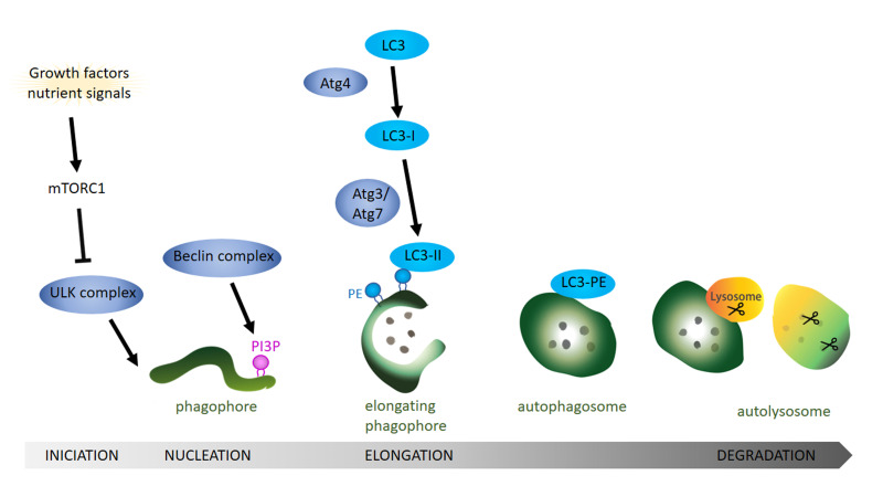
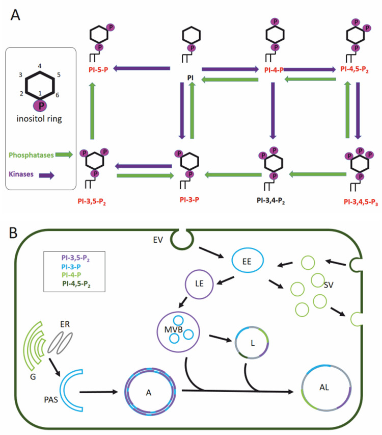
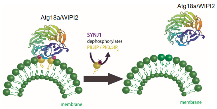
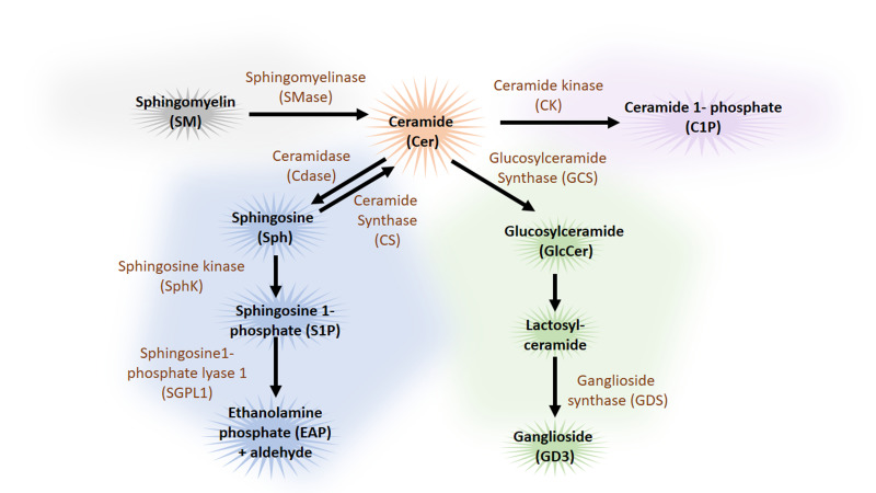
Neurodegenerative diseases are, at present, major socio-economic burdens without effective treatments and their increasing prevalence means that these diseases will be a challenge for future generations. Neurodegenerative diseases may differ in etiology and pathology but are often caused by the accumulation of dysfunctional and aggregation-prone proteins. Autophagy, a conserved cellular mechanism, deals with cellular stress and waste product build-up and has been shown to reduce the accumulation of dysfunctional proteins in animal models of neurodegenerative diseases. Historically, progress in understanding the precise function of lipids has traditionally been far behind other biological molecules (like proteins) but emerging works demonstrate the importance of lipids in the autophagy pathway and how the disturbance of lipid metabolism is connected to neurodegeneration. Here we review how altered autophagy and the disturbance of lipid metabolism, particularly of phosphoinositols and sphingolipids, feature in neurodegenerative diseases and address work from the field that suggests that these potentially offer an opportunity of therapeutic intervention.

Keywords: Alzheimer's disease; Parkinson's disease; autophagy; lipids; neurodegeneration; phosphoinositols; sphingolipids.


Conflict of interest statement

Conflict of interest: The authors declare no conflict of interest.

Figures

Figure 1
Figure 1. FIGURE 1: General overview of the autophagy pathway.
Schematic drawing showing the autophagy process starting with the formation of the phagophore, followed by the completion of the autophagosome and finishing with the fusion of the autophagosome with the lysosome. Growth factors and nutrient signals inactivate the mammalian target of rapamycin complex 1 (mTOR1) leading to the activation and recruitment of the ULK complex to the phagophore. Activity of the Beclin complex leads to local enrichment of phosphatidylinositol-3-phosphate (PI3P). During the elongation of the phagophore, ATG4 processing of LC3 (ATG8 in Drosophila) and the subsequent conjugation to phosphatidylethanolamine (PE) on the phagophore membrane via ATG3 and ATG7 is essential step to form the autophagosome. After the fusion of the lysosome with the mature autophagosome, lysosomal proteases like cathepsin D degrade the autophagosomal content.
Figure 2
Figure 2. FIGURE 2: Phosphatidylinositol pathway and its role in cellular trafficking.
(A) Phosphatidylinositols (PtdIns) are small lipids, consisting of two fatty acid chains, a glycerol backbone and an inositol ring. Kinases (violet) can phosphorylate the inositol ring at various positions (3, 4 and 5) leading to different phosphoinositides. We can differentiate between phosphatidylinositol monophosphates (PI3P, PI4P, and PI5P), diphosphates (PI3,4P2, PI3,5P2, PI4,5P2), and a triphosphate (PI3,4,5P3), that are substrates of different phosphatases (green) and kinases (see text for more details). Phosphoinositides that participate in the autophagy pathway are marked in red. (B) Membrane anchoring of the PtdIns is possible thanks to its glycerol backbone that positions the inositol ring towards the cytoplasmic site. Therefore, PtdIns often act as recognition motif for proteins containing a specific PI3P binding domain like pleckstrin homology (PH), FYVE, WD40 repeats, FERM, PTB, and PDZ to mediate signaling pathways including autophagy and clathrin-mediated endocytosis. Moreover, the specific enrichment of PtdIns can even mark the identity of intracellular organelles. Membranes are colored depending on postulated enrichment of specific phosphoinositols. PI4P enriched organelles are synaptic vesicles and Golgi, the phagophore and the early endosome are enriched in PI3P while the late endosome is enriched in PI3,5P2. The multivesicular body, a specialized late endosome, and the autophagosome are PI3,5P2 and PI3P positive organelles. Once the autophagosome fuses with the lysosome, the resulting autolysosome becomes enriched in PI3P, PI3,5P2 and PI4P. Golgi (G), endoplasmic reticulum (ER), phagophore assembly site (PAS), autophagosome (A), autolysosome (AL), multivesicular bodies (MVB), lysosome (L), endocytotic vesicle (EV), early endosome (EE), late endosome (LE), synaptic vesicle (SV).
Figure 3
Figure 3. FIGURE 3: Maturation of autophagosomes relies on the turnover of PI3P/PI3,5P2 on autophagosomal membranes.
In the initial phase of the autophagy pathway the accumulation of PI3P on the phagophore is required to recruit ATG18 and its mammalian homologue WIPI2. The WD40 domain in ATG18/WIPI2 folds into a seven bladed beta-propeller that contains two phenylalanine-arginine-arginine-glycine (FRRG) motifs in the sixth blade. This FRRG residue is a two sided recognition motif with binding specificity to PI3P/PI3,5P2. Maturation of nascent autophagosomes requires the shedding of autophagic factors like ATG18/WIPI2. At the presynaptic terminal, the phosphatase synaptojanin1 (Synj1) dephosphorylates PI3P/PI3,5P2 on autophagosomal membranes leading to the removal ATG18/WIPI2. Shedding of autophagic factors like ATG18/WIPI2 from the autophagosome is important for progression of the autophagic pathway.
Figure 4
Figure 4. FIGURE 4: Overview of sphingolipid metabolism pathway.
Ceramide (Cer) occupies a central position in the catabolic (blue), the sphingomyelin (grey) and hydrolytic (green) sphingolipid pathways. Apart from the de novo synthesis, ceramide can also be synthesized from sphingomyelin (SM) by sphingomyelinase (SMase) or from sphingosine (Sph) by ceramide synthase (CS). Cer can be catabolized to the biologically active metabolites, sphingosine (Sph) and sphingosine 1-phosphate (S1P) and further catabolized to ethanolamine phosphate (EAP) and C16 fatty aldehydes by ceramidase (Cdase) by the sphingosine kinase (SphK) and sphingosine1-phosphate lyase 1 (SGPL1) respectively. Cer can also be hydrolysed by glucosylceramide synthase (GCS) to glucosylceramide (GlcCer) and by ganglioside synthase (GDS) to ganglioside (GD3), a complex glycosphingolipid. Phosphorylation of Cer by the ceramide kinase (CK) produces ceramide-1-phosphate (C1P). Sphingolipids are coloured in black and enzymes are coloured in brown. (Note only autophagy relevant parts of the sphingolipid pathways are illustrated.)
Figure 5
Figure 5. FIGURE 5: Bioactive sphingolipids, ceramide and sphingosine-1-phosphate (S1P) regulate autophagy especially in the initial steps.
Lipids from the catabolic (blue), the sphingomyelin (grey) and hydrolytic (green) sphingolipid pathways function in the autophagy pathway. Sphingomyelin (SM) conversion to ceramide (Cer) by sphingomyelinase (SMase) affects mTORC1 activity on the lysosome and also on the phagophore. Cer can also induce autophagy by activating the transcription of ATG, BECN1 (Beclin) and LC3. Glycosphingolipids, glucosylceramide (GlcCer) and ganglioside (GD3) affect the autophagy pathway via mTORC1 or the Beclin complex respectively. LC3 conjugation to phosphatidylethanolamine (PE) is modulated by the catabolic sphingolipid pathway since sphingosine-1-phosphate (S1P) conversion to ethanolamine phosphate (EAP) that can be further converted to PE. During auto-lysosomal fusion Cer and SP1 have opposing effects (See text for details). Ceramide (Cer), sphingomyelin (SM), sphingomyelinase (SMase), acid sphingomyelinase (ASM), glucosylceramide (GlcCer), glucosylceramide synthase (GCS), ganglioside (GD3), sphingosine-1-phosphate (S1P), sphingosine-1-phosphate lyase 1 (SGPL1), ethanolamine phosphate (EAP), phosphatidylethanolamine (PE).
Figure 6
Figure 6. FIGURE 6: Proposed link of lipids in autophagy and neurodegeneration.
There is a tight relationship between lipid metabolism and autophagy. Various sphingolipids function as bioactive lipids in the regulation of autophagy, while phosphoinositide phosphates (PtdIns) mainly function in the recruitment of autophagic factors. Autophagy is known to be an essential physiological mechanism to degrade dysfunctional and aggregate-prone proteins that are commonly accumulated in neurodegenerative disease. Changes in the sphingolipid rheostat (imbalance in the ratio between the concentrations of the apoptosis activator ceramide, and the anti-apoptotic mediator sphingosine-1-phosphate) have been implicated in many neurodegenerative diseases. Deregulation of phosphatidylinositol levels lead to neurodegeneration and various phosphatases and kinases targeting phosphatidylinositols have been found to be mutated in various neurodegenerative diseases.

References

    1. Bach J-P, Ziegler U, Deuschl G, Dodel R, Doblhammer-Reiter G. Projected numbers of people with movement disorders in the years 2030 and 2050. Mov Disord. 2011;26(12):2286–2290. doi: 10.1002/mds.23878. - DOI - PubMed
    1. Dorsey ER, Constantinescu R, Thompson JP, Biglan KM, Holloway RG, Kieburtz K, Marshall FJ, Ravina BM, Schifitto G, Siderowf A, Tanner CM. Projected number of people with Parkinson disease in the most populous nations, 2005 through 2030. Neurology. 2007;68:384–386. doi: 10.1212/01.wnl.0000271777.50910.73. - DOI - PubMed
    1. Mayeux R, Stern Y. Epidemiology of Alzheimer disease. Cold Spring Harb Perspect Med. 2012;2(8):a006239. doi: 10.1101/cshperspect.a006239. - DOI - PMC - PubMed
    1. Spillantini MG, Schmidt ML, Lee VM-Y, Trojanowski JQ, Jakes R, Goedert M. α-Synuclein in Lewy bodies. Nature. 1997;388(6645):839–840. doi: 10.1038/42166. - DOI - PubMed
    1. Zhu X, Siedlak SL, Smith MA, Perry G, Chen SG. LRRK2 protein is a component of lewy bodies. Ann Neurol. 2006;60(5):617–618. doi: 10.1002/ana.20928. - DOI - PubMed

LinkOut - more resources