Case Reports
. 2020 Dec;39(12):2477-2480.
doi: 10.1007/s10096-020-03980-y. Epub 2020 Jul 12.

First report of a new corneal pathogen: Phaeoacremonium parasiticum

Affiliations
Case Reports

First report of a new corneal pathogen: Phaeoacremonium parasiticum

Horace Massa et al. Eur J Clin Microbiol Infect Dis. 2020 Dec.

Abstract

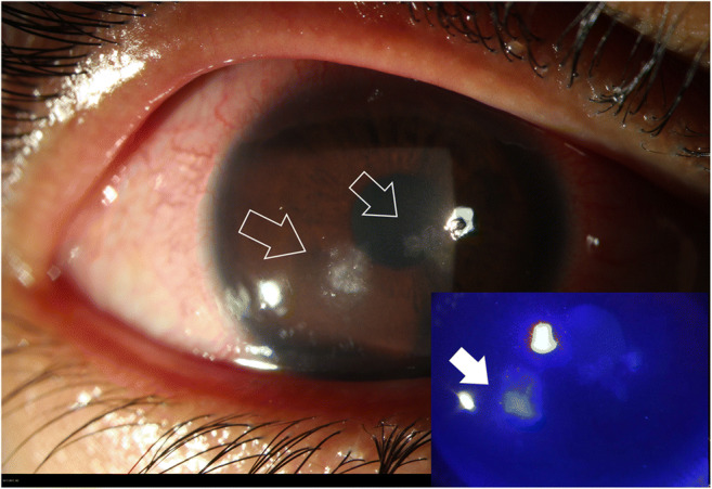
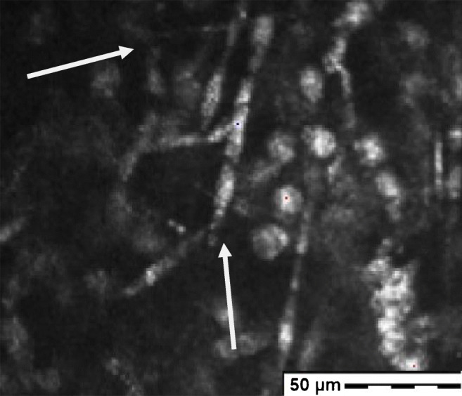
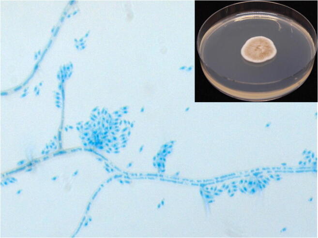
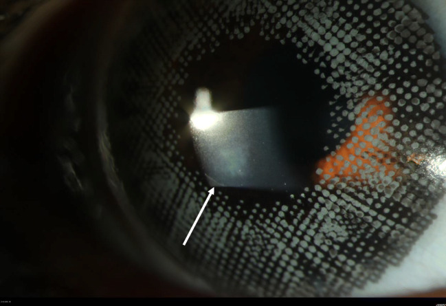
Keratitis is a public health issue in developing countries and a potentially sight-threatening condition. Collagen fibrils in the corneal stroma are parallels to each other. Fundamental substance maintains the same space between collagen fibrils. That is how corneal transparency can be achieved. Any damage which can modify this structure will lead to corneal opacity and loss of vision. Fungal keratitis might appear in up to one-third of cases. Nevertheless, fungal keratitis remains poorly described and understood. Herein, we present the first ever reported case of corneal infection due to Phaeoacremonium parasiticum in a young patient. We describe the clinical and microbial characteristics, and we also discuss the use of confocal microscopy in early diagnosis of this infection.

Keywords: Confocal in vivo imaging; Eye; Fungus; Keratitis; Microbiology; Phaeoacremonium.


Conflict of interest statement

The authors declare that they have no conflict of interest.

Figures

Fig. 1
Fig. 1
Slit lamp images of the right eye: white arrows pointing the fungal keratitis with one satellite lesion. Bottom right, the same image with fluorescein test revealing epithelium damage and the stromal impregnation of the yellowish dye (full white arrow)
Fig. 2
Fig. 2
Image of the corneal lesion showing hyper reflective round structure corresponding to inflammatory cells and the presence of highly reflective fungal hyphae (white arrows) at 70 microns stromal depth (picture acquired with the Heidelberg Retina Tomograph II Rostock Cornea Module)
Fig. 3
Fig. 3
Main picture: lactophenol cotton blue stain reveals phialides bearing apical clusters of cylindrical to sausage-shaped hyaline conidia. Top left: the colonies have a moderate growth in Sabouraud agar at 30 °C; the appearance of the colonies is velvety, white-grey to brown with radial furrows
Fig. 4
Fig. 4
Slit lamp image of the cornea with a healthy epithelium and a sub epithelial scare (white arrow) 1 year after discharging the patient. Note that the patient is still wearing cosmetic contact lenses

References

    1. Bourne RRA, Jonas JB, Bron AM, et al. Prevalence and causes of vision loss in high-income countries and in Eastern and Central Europe in 2015: magnitude, temporal trends and projections. Br J Ophthalmol. 2018;102:575–585. doi: 10.1136/bjophthalmol-2017-311258. - DOI - PMC - PubMed
    1. Hassell JR, Birk DE. The molecular basis of corneal transparency. Exp Eye Res. 2010;91:326–335. doi: 10.1016/j.exer.2010.06.021. - DOI - PMC - PubMed
    1. Chen S, Mienaltowski MJ, Birk DE. Regulation of corneal stroma extracellular matrix assembly. Exp Eye Res. 2015;133:69–80. doi: 10.1016/j.exer.2014.08.001. - DOI - PMC - PubMed
    1. AlMahmoud T, Elhanan M, Elshamsy MH, Alshamsi HN, Abu-Zidan FM. Management of infective corneal ulcers in a high-income developing country. Medicine (Baltimore) 2019;98:e18243. doi: 10.1097/MD.0000000000018243. - DOI - PMC - PubMed
    1. Menda SA, Das M, Panigrahi A et al (2019) Association of postfungal keratitis corneal scar features with visual acuity. JAMA Ophthalmol 138(2):113–118 - PMC - PubMed

Publication types

Supplementary concepts