A unique bacterial tactic to circumvent the cell death crosstalk induced by blockade of caspase-8
- PMID: 32657447
- PMCID: PMC7459423
- DOI: 10.15252/embj.2020104469
A unique bacterial tactic to circumvent the cell death crosstalk induced by blockade of caspase-8
Abstract
Upon invasive bacterial infection of colonic epithelium, host cells induce several types of cell death to eliminate pathogens. For instance, necroptosis is a RIPK-dependent lytic cell death that serves as a backup system to fully eliminate intracellular pathogens when apoptosis is inhibited; this phenomenon has been termed "cell death crosstalk". To maintain their replicative niche and multiply within cells, some enteric pathogens prevent epithelial cell death by delivering effectors via the type III secretion system. In this study, we found that Shigella hijacks host cell death crosstalk via a dual mechanism: inhibition of apoptosis by the OspC1 effector and inhibition of necroptosis by the OspD3 effector. Upon infection by Shigella, host cells recognize blockade of caspase-8 apoptosis signaling by OspC1 effector as a key danger signal and trigger necroptosis as a backup form of host defense. To counteract this backup defense, Shigella delivers the OspD3 effector, a protease, to degrade RIPK1 and RIPK3, preventing necroptosis. We believe that blockade of host cell death crosstalk by Shigella is a unique intracellular survival tactic for prolonging the bacterium's replicative niche.
Keywords: Shigella; apoptosis; effector; necroptosis.
© 2020 The Authors. Published under the terms of the CC BY 4.0 license.
Conflict of interest statement
The authors declare that they have no conflict of interest.
Figures

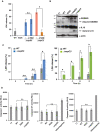
HT29 cells were infected with Shigella WT, S325, or ospD deletion mutants and incubated for 8 h. Aliquots of cellular supernatants were subjected to cytotoxicity assays. *P < 0.05 (one‐way ANOVA).
HT29 cells were infected with Shigella WT or ΔospD3 and incubated for 8 h. Infected cells were fixed and subjected to TUNEL and PI staining. Percentages of positive cells (TUNEL, green; PI, red) are shown in graph at right. The nuclei were stained with DAPI (blue). Scale bar: 100 μm. n.s., not significant; *P < 0.05 (unpaired two‐tailed Student's t‐test).
HT29 cells were infected with Shigella WT, S325, or ΔospD3 and incubated for 8 h. Infected cells were subjected to Giemsa staining. Arrows indicate cells in which the cytoplasm disappeared. Scale bar: 20 μm.

HT29 cells were infected with the indicated Shigella strains in the presence or absence of caspase inhibitor (Z‐VAD-fmk, 10 μM) and incubated for 8 h. Aliquots of cellular supernatants were subjected to cytotoxicity assays. n.s., not significant; *P < 0.05 (unpaired two‐tailed Student's t‐test).
HT29 cells were infected with the indicated Shigella strains. After 8 h of incubation, infected cells were harvested and subjected to immunoblotting.
HT29 cells were infected with Shigella WT, ΔospD3, or ΔospC3 strains. Aliquot of cellular supernatants obtained at the indicated time points were subjected to cytotoxicity assay. n.s., not significant; *P < 0.05 (unpaired two‐tailed Student's t‐test).
HT29 cells were infected with the indicated Shigella strains or stimulated with staurosporine, and then incubated for 8 h. Cells were harvested and subjected to measurement of caspase activity. n.s., not significant (one‐way ANOVA).

- A
HT29 cells were infected with Shigella WT, S325, or ospD deletion mutants and incubated for 8 h. Cell lysates were subjected to immunoblotting.
- B, C
HT29 cells were infected with the indicated Shigella strains in the presence or absence of RIPK1 inhibitor, RIPK3 inhibitor, or caspase inhibitor (z‐VAD) and then incubated for 8 h. Cell lysates and aliquots of cellular supernatants were subjected to immunoblotting (B) and cytotoxicity assay (C), respectively. *P < 0.05, n.s., not significant (one‐way ANOVA).
- D, E
HT29 cells treated with the indicated siRNAs were infected with Shigella WT or ΔospD3. Cell lysates and aliquots of cellular supernatants were subjected to immunoblotting (top) and cytotoxicity assays (bottom), respectively. The knockdown efficiency of the indicated siRNAs was assessed by immunoblotting (inset). n.s., not significant; *P < 0.05 (unpaired two‐tailed Student's t‐test).

- A
HT29, HeLa, HCT116, and HaCaT cells were infected with Shigella WT, S325, or ospD3, and then incubated at 37°C for 8 h. Aliquots of cellular supernatants were subjected to cytotoxicity assays. n.s., not significant; *P < 0.05 (unpaired two‐tailed Student's t‐test).
- B
Infected cells were harvested and subjected to immunoblotting.
- C
Cell lysates of each cell line were subjected to immunoblotting.
- D, E
HeLa cells stably expressing GFP or RIPK3 were infected with Shigella WT or ΔospD3 and then incubated for 8 h. Cell lysates and aliquots of cellular supernatants were subjected to immunoblotting (D) and cytotoxicity assay (E), respectively. *P < 0.05; n.s., not significant (unpaired two‐tailed Student's t‐test).

- A
HT29 cells were infected with the indicated Shigella strains and incubated for 8 h, and then, cell lysates were subjected to immunoblotting.
- B
293T cells were transfected with Myc‐tagged ospD expression plasmids. After 24 h, cells were harvested and subjected to immunoblotting. Arrows indicate cleaved RIPK1.
- C
Multiple sequence alignment of the OspD family: Shigella OspD3, EPEC EspL, and EHEC EspL2. Conserved amino acids are indicated by asterisks. Typical protease catalytic sites are colored in red.
- D
293T cells were transfected with a series of plasmids expressing ospD3 point mutants. After 24 h, cells were harvested and subjected to immunoblotting. Arrows indicate cleaved RIPK1.
- E, F
HT29 cells were infected with Shigella WT, S325, ΔospD3, ΔospD3/D3 (ΔospD3 complemented with wild‐type ospD3), or ΔospD3/D3CS (ΔospD3 complemented with a protease activity‐deficient mutant, in which the cysteine residue at position 64 was replaced by serine) strains and incubated for 8 h. Cell lysates and aliquots of cellular supernatants were subjected to immunoblotting (E) and cytotoxicity assays (F), respectively. *P < 0.05 (one‐way ANOVA).

HT29 cells were infected with the indicated Shigella, EPEC, or EHEC strains. After 8 h of incubation, infected cells were harvested and subjected to immunoblotting.
HT29 cells were infected with the indicated Shigella, EPEC, or EHEC strains for 8 h. Aliquots of cellular supernatants were subjected to cytotoxicity assays. *P < 0.05; n.s., not significant (unpaired two‐tailed Student's t‐test).
293T cells were transfected with a series of plasmids expressing point mutants of ospD homologs (the cysteine residue at position 64 of Shigella ospD3 was replaced by alanine). After 24 h, the cells were harvested and subjected to immunoblotting. Arrows indicate cleaved RIPK1.

293T cells were transfected with a series of RIPK1 truncations, along with plasmids expressing OspD3 or OspD3‐CS (protease activity‐deficient mutant, in which the cysteine residue at position 64 was replaced by serine). After 24 h, cells were harvested and subjected to immunoblotting. Asterisks indicate the non‐cleaved form of truncated RIPK1.
Sequence alignment of the RIPK1 and RIPK3 (top). 293T cells were transfected with a series of plasmids expressing the RIPK1 RHIM domain mutant (4A and 3A indicate alanine replacement of four and three amino acids, respectively, within the indicated regions of RIPK1) along with OspD3 or OspD3‐CS. After 24 h, cells were harvested and subjected to immunoblotting (bottom).
293T cells were transfected with plasmids expressing RIPK1, RIPK1 RHIM mutant, RIPK3, or RIPK3 RHIM mutant along with empty vector, OspD3, or OspD3‐CS. After 24 h, cells were harvested and subjected to immunoblotting.
HeLa cells stably expressing RIPK3 or RIPK3‐4A were infected with Shigella WT or ΔospD3, and incubated for 8 h. Cell lysates were subjected to immunoblotting.

HT29 cells were infected with the indicated Shigella strains and incubated for 8 h. Cells were harvested and subjected to measurement of caspase‐8 activation. Caspase‐8 activity is reported as relative light units (RLU) of infected samples, minus the value in uninfected samples. *P < 0.05 (one‐way ANOVA).
HT29 cells were infected with the indicated Shigella strains and incubated for 8 h. Cell lysates were subjected to immunoblotting. Arrows indicate cleaved forms of caspase‐8, PARP, GSDMD, and IL‐18, respectively.
HT29 cells were infected with the indicated Shigella strains and incubated for 8 h. Aliquots of cellular supernatants were subjected to cytotoxicity assay. *P < 0.05; n.s., not significant (one‐way ANOVA).
HT29 cells were infected with Shigella WT or ΔospC1 mutant and incubated for 8 h. Infected cells were then fixed and stained with cleaved caspase‐8 (green), rhodamine–phalloidin (red), and DAPI (blue). Percentages of positive cells are shown in the graph at right (*P < 0.05; unpaired two‐tailed Student's t‐test). Scale bar: 100 μm.
HT29 cells were infected with the indicated Shigella strains and incubated for 8 h. Cells were harvested and subjected to measurement of caspase‐3/7 activation. Caspase‐3/7 activity is reported as relative light units (RLU) of infected samples minus uninfected samples. *P < 0.05 (one‐way ANOVA).
HT29 cells were infected with Shigella WT, S325, or ΔospC1 strains and incubated for up to 8 h. After incubation for the indicated times, cells were subjected to real‐time Annexin V apoptosis assay. Annexin V binding is reported as relative light units (RLU) in infected samples minus the value in uninfected samples. *P < 0.05 (one‐way ANOVA).

- A, B
HT29 cells were infected with Shigella WT, ΔospC1, ΔospD3, ΔospC1ΔospD3, or ΔospC1ΔospD3/ospC1 (ΔospC1ΔospD3 complemented with ospC1) strains and incubated for 8 h. Cell lysates and aliquots of cellular supernatants were subjected to immunoblotting (A) and cytotoxicity assay (B), respectively. *P < 0.05; n.s., not significant (one‐way ANOVA).
- C
HT29 cells were infected with Shigella WT, ΔospD3, or ΔospC1. Cell lysates obtained at the indicated time points were subjected to immunoblotting.
- D, E
HT29 cells treated with DMSO or caspase‐8 inhibitor were infected with Shigella WT, ΔospC1, ΔospD3, or ΔospC1ΔospD3 strains and incubated for 8 h. Cell lysates and aliquots of cellular supernatants were subjected to immunoblotting (D) and cytotoxicity assay (E), respectively. *P < 0.05; n.s., not significant (unpaired two‐tailed Student's t‐test).
- F
HT29 cells treated with the control or caspase‐8 siRNAs were infected with the indicated Shigella strains and incubated for 8 h. Cell lysates were subjected to immunoblotting. The knockdown efficiency of the indicated siRNAs was assessed by immunoblotting.

HT29 cells were infected with the indicated Shigella strains and incubated for 8 h. Cell lysates were subjected to immunoblotting.
HT29 cells were infected with Shigella WT, ΔospD3, or ΔospC1 strains. Cell lysates obtained at the indicated time points were subjected to immunoblotting.
HT29 cells were infected with Shigella WT or ΔospC3 strains. Cell lysates obtained at the indicated time points were subjected to immunoblotting.
HT29 cells treated with control or caspase‐8 siRNAs were infected with the indicated Shigella strains and incubated for 8 h. Aliquots of cellular supernatants were subjected to cytotoxicity assay. *P < 0.05; n.s., not significant (unpaired two‐tailed Student's t‐test).

Comment in
-
Manipulation of epithelial cell death pathways by Shigella.EMBO J. 2020 Sep 1;39(17):e106202. doi: 10.15252/embj.2020106202. Epub 2020 Aug 10. EMBO J. 2020. PMID: 32869315 Free PMC article.
References
-
- Ashida H, Toyotome T, Nagai T, Sasakawa C (2007) Shigella chromosomal IpaH proteins are secreted via the type III secretion system and act as effectors. Mol Microbiol 63: 680–693 - PubMed
-
- Ashida H, Kim M, Sasakawa C (2014) Manipulation of the host cell death pathway by Shigella . Cell Microbiol 16: 1757–1766 - PubMed
Publication types
MeSH terms
Substances
Grants and funding
LinkOut - more resources
Full Text Sources
Molecular Biology Databases
Miscellaneous

