. 2020 Jun 23:(160):10.3791/61392.
doi: 10.3791/61392.

Monitoring Cancer Cell Invasion and T-Cell Cytotoxicity in 3D Culture

Affiliations

Monitoring Cancer Cell Invasion and T-Cell Cytotoxicity in 3D Culture

Yuan-Na Lin et al. J Vis Exp. .

Abstract

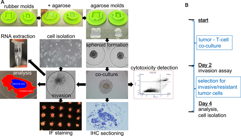
Significant progress has been made in treating cancer with immunotherapy, although a large number of cancers remain resistant to treatment. A limited number of assays allow for direct monitoring and mechanistic insights into the interactions between tumor and immune cells, amongst which, T-cells play a significant role in executing the cytotoxic response of the adaptive immune system to cancer cells. Most assays are based on two-dimensional (2D) co-culture of cells due to the relative ease of use but with limited representation of the invasive growth phenotype, one of the hallmarks of cancer cells. Current three-dimensional (3D) co-culture systems either require special equipment or separate monitoring for invasion of co-cultured cancer cells and interacting T-cells. Here we describe an approach to simultaneously monitor the invasive behavior in 3D of cancer cell spheroids and T-cell cytotoxicity in co-culture. Spheroid formation is driven by enhanced cell-cell interactions in scaffold-free agarose microwell casts with U-shaped bottoms. Both T-cell co-culture and cancer cell invasion into type I collagen matrix are performed within the microwells of the agarose casts without the need to transfer the cells, thus maintaining an intact 3D co-culture system throughout the assay. The collagen matrix can be separated from the agarose cast, allowing for immunofluorescence (IF) staining and for confocal imaging of cells. Also, cells can be isolated for further growth or subjected to analyses such as for gene expression or fluorescence activated cell sorting (FACS). Finally, the 3D co-culture can be analyzed by immunohistochemistry (IHC) after embedding and sectioning. Possible modifications of the assay include altered compositions of the extracellular matrix (ECM) as well as the inclusion of different stromal or immune cells with the cancer cells.


Conflict of interest statement

DISCLOSURES:

The authors declare that they have no competing financial interests.

Figures

Figure 1:
Figure 1:. Workflow, analyses and timeline of experiments.
(A) An 81-microwell and 35-microwell rubber mold are filled with 2% agarose in 1X PBS to generate an agarose cast with multiple microwells. Spheroids are formed upon cell seeding into the chambers of the agarose cast in a single pipetting step. Co-culture with T-cells is performed within the same cast. Functional monitoring and potential assays are shown. (B) Timeline of experiments. Tumor cells are co-cultured with autologous T-cells for 2 days allowing for a maximum interaction between both cell types. The invasion assay is initiated after two days. Endpoint analyses are performed after further two days to monitor the invasive and survival phenotype of tumor cells as well as the proliferation and survival of T-cells.
Figure 2:
Figure 2:. Quantification of invasion.
Invasion can be quantified by image analysis, e.g. using Image J software and counting the number of “spikes” per spheroid. (A) Calculation of the total invasion area as a ratio of total area to the spheroid area. (B) Examples of two different primary murine pancreatic cancer cell lines (cell line 1 and 2) in spheroid formation at different magnifications and invasion into type I collagen. (C) Analysis of invasion is performed by counting the number of spikes per spheroid (left diagram) and calculate the invasion area as described (right diagram).
Figure 3:
Figure 3:. Co-culture with dye-labeled tumor and T-cells.
(A-E) Mix of two differently dye-labeled primary murine pancreatic cancer clonal cell lines (green and red) and magnified view of one representative microwell (B-E). (F-J) Co-culture of pre-labeled tumor (green) and T-cells (red). (G-J) Magnified view of one representative microwell shows one tumor – T-cell co-culture upon tumor spheroid formation.
Figure 4:
Figure 4:. Immunofluorescence staining.
Immunofluorescence (IF) staining was performed after separating the collagen matrix from the agarose cast. (A-B) After IF staining, the collagen patches are transferred to a glass slide and covered with glass coverslips. (C-F) Example of an IF-stained collagen patch including tumor spheroids with (C) Hoechst, (D) keratin 8, (E) phalloidin and (F) overlay. G-J show the respective magnified view of a single spheroid. Bar: 300 μm.
Figure 5:
Figure 5:. Immunohistochemistry sectioning.
The agarose cast with the 3D culture immersed in Hydroxyethyl agarose processing gel, was embedded in paraffin, sectioned and processed for immunohistochemistry (IHC) staining. (A) Paraffin block used for horizontal sectioning that starts from the bottom of the agarose cast to obtain serial sections of multiple tumor cell / T-cell co-cultures within a single cast. (B) H&E – stained section of an agarose cast containing 3D co-culture of tumor and T-cells. (C) Magnified view of an H&E – stained co-culture within the agarose cast. CD8 staining of T-cells co-cultured with sensitive (D) and resistant (E) tumor cells.

References

    1. Chen DS & Mellman I Elements of cancer immunity and the cancer-immune set point. Nature 541 (7637), 321–330, doi: 10.1038/nature21349 (2017). - DOI - PubMed
    1. Sharma P & Allison JP Dissecting the mechanisms of immune checkpoint therapy. Nature Reviews Immunology 20 (2), 75–76, doi: 10.1038/s41577-020-0275-8 (2020). - DOI - PubMed
    1. Blomberg OS, Spagnuolo L, de Visser KE Immune regulation of metastasis: mechanistic insights and therapeutic opportunities. Disease Model Mechanisms 11 (10), doi: 10.1242/dmm.036236 (2018). - DOI - PMC - PubMed
    1. Schmeichel KL & Bissell MJ Modeling tissue-specific signaling and organ function in three dimensions. Journal of Cell Science 116 (12), 2377–2388, doi:10.1242/jcs.00503 (2003). - DOI - PMC - PubMed
    1. Kramer N, et al.In vitro cell migration and invasion assays. Mutation Research 752 (1), 10–24, doi: 10.1016/j.mrrev.2012.08.001 (2013). - DOI - PubMed

Publication types

Substances