Review
. 2021 Mar;76(3):698-713.
doi: 10.1111/all.14498. Epub 2020 Aug 12.

SARS-CoV-2, COVID-19, skin and immunology - What do we know so far?

Affiliations
Review

SARS-CoV-2, COVID-19, skin and immunology - What do we know so far?

Natalijia Novak et al. Allergy. 2021 Mar.

Abstract

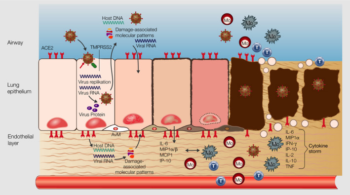
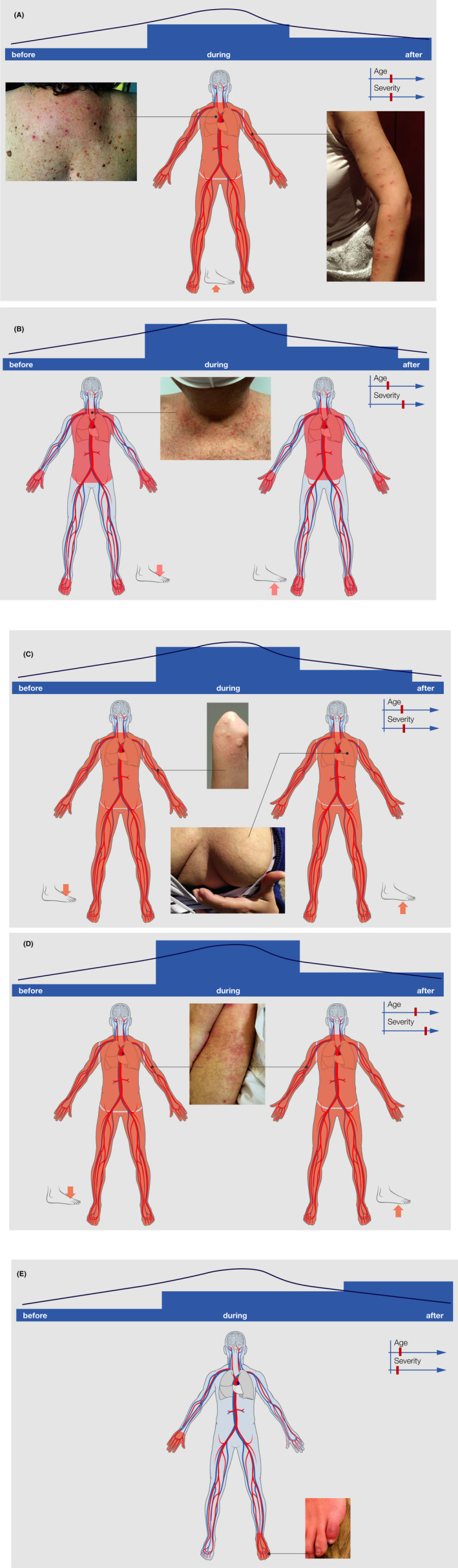
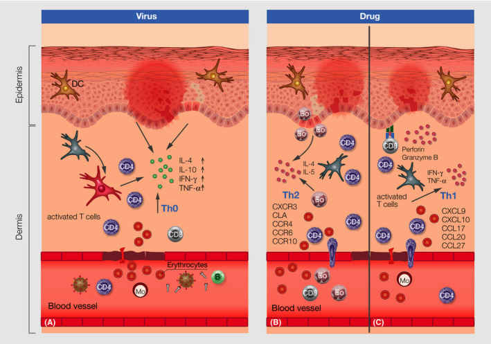
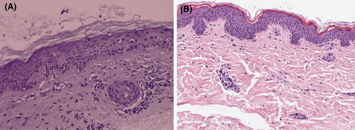
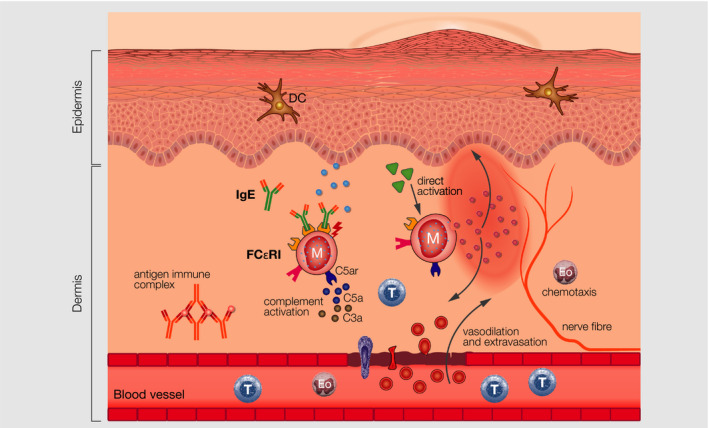
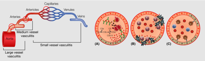
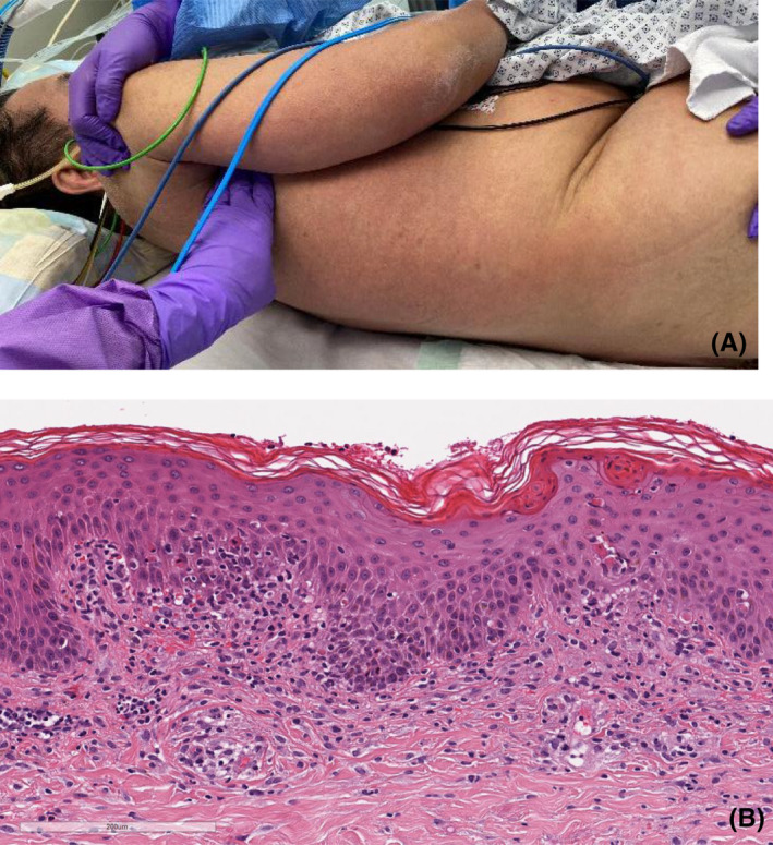
The pandemic condition coronavirus disease (COVID-19), caused by the novel severe acute respiratory syndrome coronavirus 2 (SARS-CoV-2), can take asymptomatic, mild, moderate, and severe courses. COVID-19 affects primarily the respiratory airways leading to dry cough, fever, myalgia, headache, fatigue, and diarrhea and can end up in interstitial pneumonia and severe respiratory failure. Reports about the manifestation of various skin lesions and lesions of the vascular system in some subgroups of SARS-CoV-2-positive patients as such features outside the respiratory sphere, are rapidly emerging. Vesicular, urticarial, and maculopapular eruptions and livedo, necrosis, and other vasculitis forms have been reported most frequently in association with SARS-CoV-2 infection. In order to update information gained, we provide a systematic overview of the skin lesions described in COVID-19 patients, discuss potential causative factors, and describe differential diagnostic evaluations. Moreover, we summarize current knowledge about immunologic, clinical, and histologic features of virus- and drug-induced lesions of the skin and changes to the vascular system in order to transfer this knowledge to potential mechanisms induced by SARS-CoV-2.

Keywords: allergy; drug hypersensitivity; skin; virus.


Conflict of interest statement

The authors declare no conflict of interest.

Figures

FIGURE 1
FIGURE 1
Structure of SARS‐CoV‐2 virus with its typical capsid and the spikes, “corona.” ssRNA, single‐stranded RNA; ACE2, angiotensin receptor 2; TMPRSS2, transmembrane protease serine subtype 2
FIGURE 2
FIGURE 2
Cascade of events in lung epithelium during SARS‐CoV‐2 infection. SARS‐CoV‐2 infects lung epithelial cells via ACE2 and TMPRSS2 receptor, the host cells undergo apoptosis in consequence of virus replication and release, and the undergoing cell releases damage‐associated molecular patterns, which induce the production of pro‐inflammatory mediators by epithelial and endothelial cells in the neighborhood. Inflammatory cells such as monocytes, macrophages, and T cells are recruited from the blood to the lung epithel, increase the production of inflammatory mediates and further infiltration of the lung by inflammatory cells, leading to damage of the lung structure and a “cytokine storm,” which reaches the vasculature and other organs. ssRNA, single‐stranded RNA; ACE2, angiotensin receptor 2; TMPRSS2, transmembrane protease serine subtype 2; Mo, monocyte, T, T lymphocyte, M, macrophage
FIGURE 3
FIGURE 3
Symptoms of COVID‐19
FIGURE 4
FIGURE 4
A: Typical skin lesions (vesicular eruptions occurring early during COVID‐19 (A), with symptoms of COVID‐19, that is, maculopapular exanthema (B), urticarial rash (C), vasculitis (D), and later during COVID‐19 disease Chilblain eruptions (E). The bar on top indicates the onset of skin symptoms in relation to COVID‐19 symptoms, as well as average age and severity of COVID‐19 patients, and typical predilection sites of the lesions are also depicted
FIGURE 5
FIGURE 5
Immunologic features in the skin of virus‐induced maculopapular exanthema (A) with virus antibody activation and interaction with immune cells in the blood, recruitment of activated cells to the skin, extravasation of erythrocytes through the blood vessels, cytokine production and keratinocyte apoptosis and related changes to the dermis and epidermis and drug‐induced maculopapular rush type IVb (B) and IVc (C) derived by activated T cells, extravasation of erythrocytes, perivascular infiltrates and perforin and granzyme B and Th1 cytokines as well as different chemokines and recruitment of T cells and eosinophils to the dermis and respective changes to the dermis and epidermis; DC, dendritic cell; B, B cell; Mo, monocyte, Eo, eosinophil
FIGURE 6
FIGURE 6
Histologic features of skin biopsies taken from two SARS‐CoV‐2‐positive patients with maculopapular eruptions. (A Skin sections showing epidermis with mild hyperkeratosis, keratinocytes with frosted glass nuclei, with intranuclear and occasionally multinucleate inclusions, reminiscent of cytopathic damage. Dermis without edema, perivascular inflammatory infiltrate extending focally to the basal layer, causing slight vacuolate damage and pigmentary incontinence. No eosinophils are observed. (B) Histology (H&E stain) showing an inconspicuous epidermis and a very subtle perivascular lymphohistiocytic infiltrate in the upper dermis with admixture of few eosinophilic granulocytes
FIGURE 7
FIGURE 7
IgE‐, direct‐, and antigen‐immunocomplex–mediated mast cell activation in urticaria
FIGURE 8
FIGURE 8
Vasculitis of small, medium, and large vessels, with formation of antigen‐immune complexes, which accumulate within the vessels and damage of the endothel, which leads to extravasation of cells and neutrophil recruitment
FIGURE 9
FIGURE 9
(A) 58‐year‐old male patient diagnosed with COVID‐19 and DRESS syndrome. (B) Histology (H&E stain) showing interface changes (vacuolar degeneration of the basal layer, apoptotic keratinocytes, exocytosis of lymphocytes) and spongiotic changes with hyperparakeratosis. Perivascular lymphohistiocytic infiltrate with admixture of few eosinophilic granulocytes. Mild extravasation of erythrocytes

References

    1. Tay MZ, Poh CM, Rénia L, MacAry PA, Ng LFP. The trinity of COVID‐19: immunity, inflammation and intervention. Nat Rev Immunol. 2020;20(6):363‐374. 10.1038/s41577-020-0311-8 - DOI - PMC - PubMed
    1. Zhu NA, Zhang D, Wang W, et al. A novel coronavirus from patients with pneumonia in China, 2019. N Engl J Med. 2020;382:727‐733. - PMC - PubMed
    1. Dong X, Cao Y‐Y, Lu X‐X, et al. Eleven faces of coronavirus disease 2019. Allergy 2020;75(7):1699‐1709. 10.1111/all.14289 - DOI - PMC - PubMed
    1. Li L‐Q, Huang T, Wang Y‐Q, et al. COVID‐19 patients' clinical characteristics, discharge rate, and fatality rate of meta‐analysis. J Med Virol. 2020;92(6):577‐583. 10.1002/jmv.25757 - DOI - PMC - PubMed
    1. Andersen KG, Rambaut A, Lipkin WI, Holmes EC, Garry RF. The proximal origin of SARS‐CoV‐2. Nat Med. 2020;26(4):450‐452. - PMC - PubMed

Publication types

Substances