. 2020 Dec 1;21(23):3417-3422.
doi: 10.1002/cbic.202000457. Epub 2020 Aug 7.

Multiparameter Kinetic Analysis for Covalent Fragment Optimization by Using Quantitative Irreversible Tethering (qIT)

Affiliations

Multiparameter Kinetic Analysis for Covalent Fragment Optimization by Using Quantitative Irreversible Tethering (qIT)

Gregory B Craven et al. Chembiochem. .

Abstract

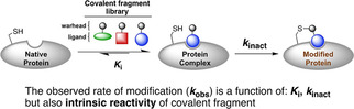
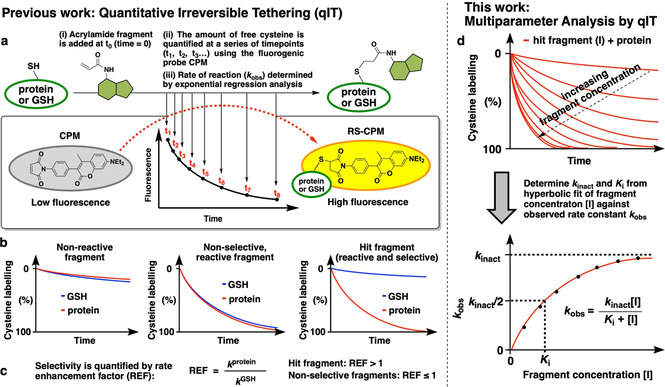
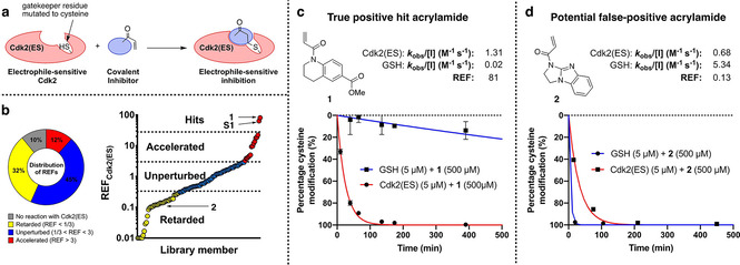
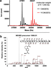
Chemical probes that covalently modify cysteine residues in a protein-specific manner are valuable tools for biological investigations. Covalent fragments are increasingly implemented as probe starting points, but the complex relationship between fragment structure and binding kinetics makes covalent fragment optimization uniquely challenging. We describe a new technique in covalent probe discovery that enables data-driven optimization of covalent fragment potency and selectivity. This platform extends beyond the existing repertoire of methods for identifying covalent fragment hits by facilitating rapid multiparameter kinetic analysis of covalent structure-activity relationships through the simultaneous determination of Ki , kinact and intrinsic reactivity. By applying this approach to develop novel probes against electrophile-sensitive kinases, we showcase the utility of the platform in hit identification and highlight how multiparameter kinetic analysis enabled a successful fragment-merging strategy.

Keywords: Cdk2; covalent fragments; covalent inhibition kinetics; electrophile-sensitive inhibition; fragment-based drug discovery.


Figures

Figure 1
Figure 1
Two‐step mechanism for covalent protein inhibition.
Figure 2
Figure 2
Comparison of previous and current applications of Quantitative irreversible tethering (qIT). a) Overview of rate determination. A cysteine‐containing biomolecule is treated with covalent fragments under pseudo‐first‐order conditions. Reaction progress is followed by discrete measurement of free cysteine concentration by using the fluorogenic probe CPM, and rate constants (k obs) are derived from exponential regression analysis. b) Comparison of the reactivity profile of each fragment with the POI and GSH typically classes them as unreactive, reactive but non‐selective or reactive and selective. c) Quantification of kinetic POI/GSH selectivity facilitates hit identification. d) The observed rate of reaction between the POI and hit fragment is determined at a range of ligand concentrations by using qIT. Subsequent hyperbolic regression analysis is used to derive K i and k inact to facilitate comprehensive covSAR analysis.
Figure 3
Figure 3
Screening cascade to identify covalent fragments targeting Cdk2(ES). a) Gatekeeper cysteine (F80C) ES strategy for allele‐specific inhibition of Cdk2. b) Distribution of rate‐enhancement factors for the covalent fragment library screened against Cdk2(ES) and GSH, highlighting hits 1 and S1 and potential false positive 2. c), d) qIT data for acrylamides 1 and 2 (0.5 mM) in reaction with Cdk2(ES) or glutathione (5 μM). Fluorescence intensity is converted into percentage cysteine modification by normalizing to DMSO control=0 %, no thiol=100 %.
Figure 4
Figure 4
a) Intact‐protein mass spectrum of Cdk2(ES) before and after incubation with acrylamide 1 (0.5 mM) for 2 h showing complete mono‐modification (ΔMW=246 Da). b) MALDI‐TOF/TOF spectrum of the C80‐containing tryptic peptide (precursor ion m/z 1866) of 1–Cdk2(ES).
Figure 5
Figure 5
In vitro kinase activity for pCdk2(WT/ES)/cyclin A2 holoenzme, demonstrating complete inhibition of pCdk2(ES) by treatment with 0.5 mM acrylamide 1 for 2 h and then subsequent dialysis to remove excess ligand: The holoenzymes were then incubated with peptide substrate, ATP, NADH, PEP, LD and PK at 37 °C, and the absorbance was measured over time in clear 384‐well plates.
Figure 6
Figure 6
Structure‐guided ligand optimization. a) Crystal structure of the 1‐Cdk2(ES) conjugate (resolution: 1.77 Å, PDB ID: 5OSM). The 2F oF c electron‐density map (blue) is contoured at 1σ around Cys80 (yellow) and the ligand (green), with the hydrogen bonds to L83 and K33 shown in red. b) Covalent SAR for fragment analogues by REF analysis. c) qIT data for acrylamide 6 (0.5 mM) in reaction with Cdk2(ES) or glutathione (5 μM). Fluorescence intensity is converted into percentage cysteine modification by normalizing to DMSO control=0 %, no thiol=100 %. d) Crystal structure of the 6‐Cdk2(ES) complex (cyan; resolution 1.72 Å, PDB ID: 5OO3) aligned against 1–Cdk2(ES) (green).
Figure 7
Figure 7
Kinetic analysis of merged fragments. a) Kinetic analysis of acrylamides 1, 6, 9, 10 and 11 determined by qIT. Concnetration‐dependent analysis was used to derive K i and k inact. b) Crystal structure of the 11‐Cdk2(ES) conjugate (resolution: 1.66 Å, PDB ID: 6YL1). The 2F oF c electron density map (blue) is contoured at 1σ around Cys80 (yellow) and the ligand (green), with the hydrogen bonds to L83 shown in red. c) Concentration‐dependent qIT analysis. Left: Representative qIT data: acrylamide 11 (25–500 μM) reacting with Cdk(ES) (5 μM; n=2, error bars denote SD). Fluorescence intensity is converted into percentage cysteine modification by normalizing to DMSO control=0 %, no thiol=100 %. Right: Hyberbolic fitting of k obs against concentration of acrylamides yields K i and k inact. d) Overlay of crystal binding poses of Cys80 conjugated to acrylamides 1, 6, 9 and 11, after alignment of the global protein structre.

References

    1. Resnick E., Bradley A., Gan J., Douangamath A., Krojer T., Sethi R., Geurink P. P., Aimon A., Amitai G., Bellini D., et al., J. Am. Chem. Soc. 2019, 141, 8951–8968. - PMC - PubMed
    1. Backus K. M., Correia B. E., Lum K. M., Forli S., Horning B. D., González-Páez G. E., Chatterjee S., Lanning B. R., Teijaro J. R., Olson A. J., et al., Nature 2016, 534, 570–574. - PMC - PubMed
    1. Erlanson D. A., Wells J. A., Braisted A. C., Annu. Rev. Biophys. Biomol. Struct. 2004, 33, 199–223. - PubMed
    1. Ostrem J. M., Peters U., Sos M. L., Wells J. A., Shokat K. M., Nature 2013, 503, 548–551. - PMC - PubMed
    1. Cravatt B. F., Wright A. T., Kozarich J. W., Annu. Rev. Biochem. 2008, 77, 383–414. - PubMed

Publication types