

Proapoptotic Peptide Brush Polymer Nanoparticles via Photoinitiated Polymerization-Induced Self-Assembly
- PMID: 32659039
- PMCID: PMC7722202
- DOI: 10.1002/anie.202006385
Proapoptotic Peptide Brush Polymer Nanoparticles via Photoinitiated Polymerization-Induced Self-Assembly
Abstract
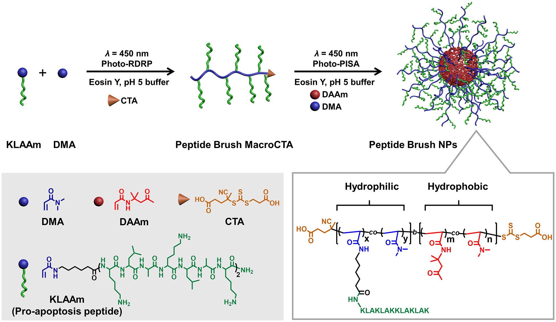
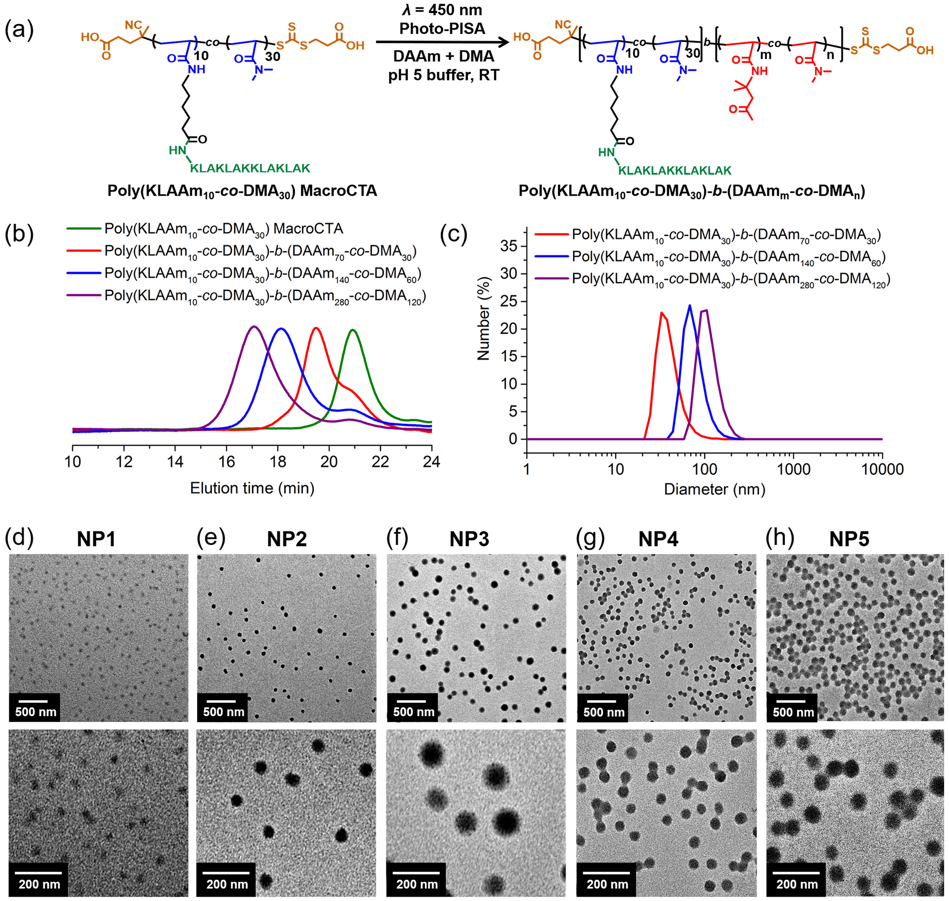
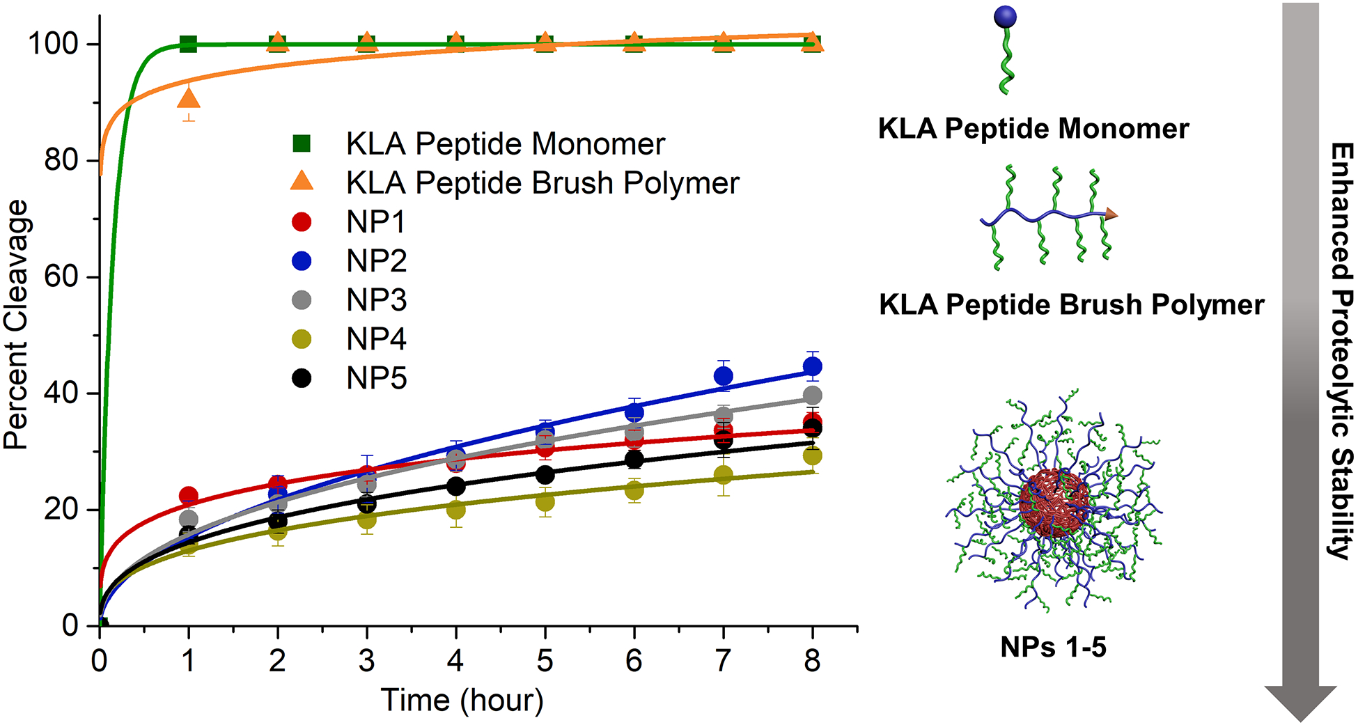
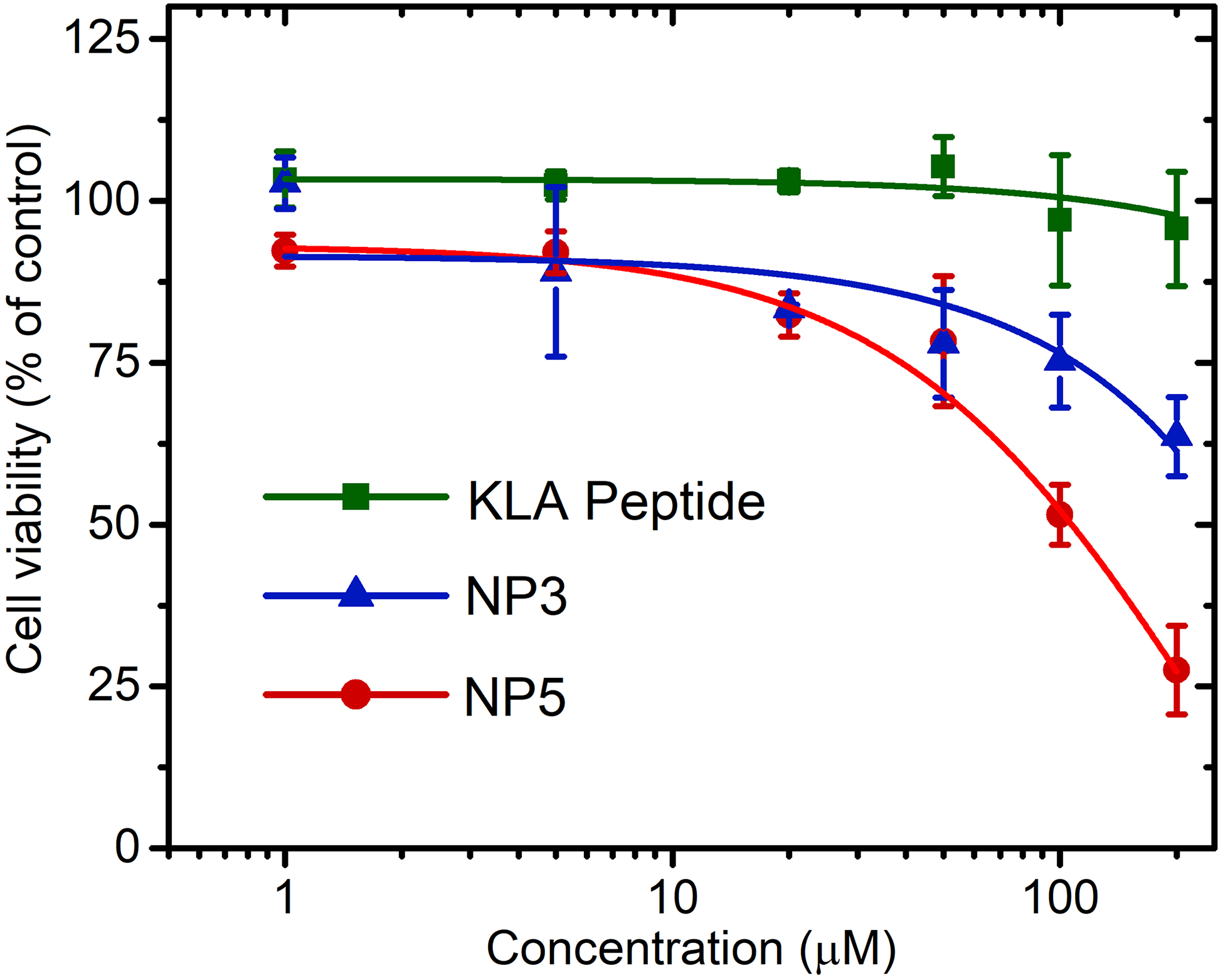
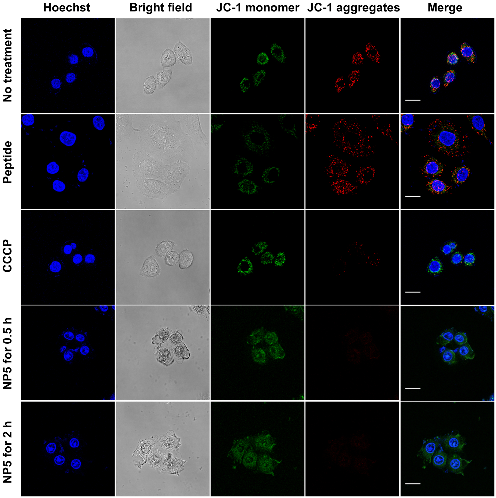
Herein, we report the photoinitiated polymerization-induced self-assembly (photo-PISA) of spherical micelles consisting of proapoptotic peptide-polymer amphiphiles. The one-pot synthetic approach yielded micellar nanoparticles at high concentrations and at scale (150 mg mL-1 ) with tunable peptide loadings up to 48 wt. %. The size of the micellar nanoparticles was tuned by varying the lengths of hydrophobic and hydrophilic building blocks. Critically, the peptide-functionalized nanoparticles imbued the proapoptotic "KLA" peptides (amino acid sequence: KLAKLAKKLAKLAK) with two key properties otherwise not inherent to the sequence: 1) proteolytic resistance compared to the oligopeptide alone; 2) significantly enhanced cell uptake by multivalent display of KLA peptide brushes. The result was demonstrated improved apoptosis efficiency in HeLa cells. These results highlight the potential of photo-PISA in the large-scale synthesis of functional, proteolytically resistant peptide-polymer conjugates for intracellular delivery.
Keywords: nanoparticles; peptide delivery; peptide-polymer conjugates; polymerization; scaled synthesis.
© 2020 Wiley-VCH GmbH.
Figures
References
-
- Yu B, Hwang D, Jeon H, Kim H, Lee Y, Keum H, Kim J, Lee DY, Kim Y, Chung J, Jon S, Angew. Chem. Int. Ed 2019, 58, 2005–2010. - PubMed
-
- Drucker DJ, Nat. Rev. Drug Discov 2020, 19, 277–289. - PubMed
-
- Navaratna T, Atangcho L, Mahajan M, Subramanian V, Case M, Min A, Tresnak D, Thurber GM, J. Am. Chem. Soc 2020, 142, 1882–1894; - PMC - PubMed
- Kasper MA, Glanz M, Oder A, Schmieder P, von Kries JP, Hackenberger CPR, Chem Sci 2019, 10, 6322–6329; - PMC - PubMed
- Menacho-Melgar R, Decker JS, Hennigan JN, Lynch MD, J. Control Release 2019, 295, 1–12; - PMC - PubMed
- Roberts MJ, Bentley MD, Harris JM, Adv. Drug Deliv. Rev 2012, 64, 116–127; - PubMed
- Webber MJ, Appel EA, Vinciguerra B, Cortinas AB, Thapa LS, Jhunjhunwala S, Isaacs L, Langer R, Anderson DG, Proc. Natl. Acad. Sci. USA 2016, 113, 14189–14194; - PMC - PubMed
- Gentilucci L, De Marco R, Cerisoli L, Curr. Pharm. Design 2010, 16, 3185–3203; - PubMed
- Chatterjee J, Gilon C, Hoffman A, Kessler H, Accounts Chem. Res 2008, 41, 1331–1342; - PubMed
- Itoh H, Miura K, Kamiya K, Yamashita T, Inoue M, Angew. Chem. Int. Ed 2020, 59, 4564–4571; - PubMed
- Wu YT, Villa F, Maman J, Lau YH, Dobnikar L, Simon AC, Labib K, Spring DR, Pellegrini L, Angew. Chem. Int. Ed 2017, 56, 12866–12872. - PubMed
-
- Guidotti G, Brambilla L, Rossi D, Trends Pharmacol. Sci 2017, 38, 406–424. - PubMed
Publication types
MeSH terms
Substances
Grants and funding
LinkOut - more resources
Full Text Sources
