

Identification of enterobacteria in viscera of pigs afflicted with porcine reproductive and respiratory syndrome and other viral co-infections
- PMID: 32659314
- PMCID: PMC7352111
- DOI: 10.1016/j.micpath.2020.104385
Identification of enterobacteria in viscera of pigs afflicted with porcine reproductive and respiratory syndrome and other viral co-infections
Abstract
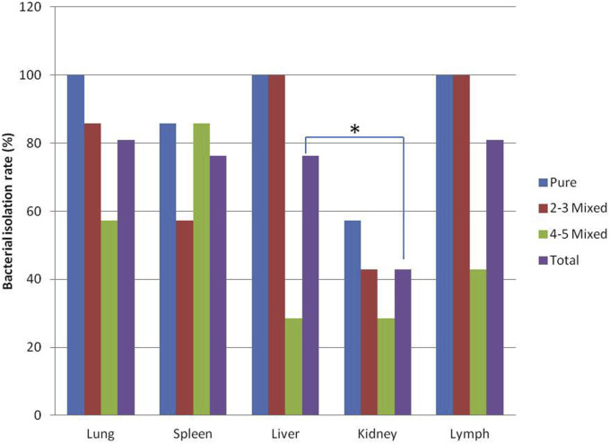
In order to investigate enterobacteria presence involved in the secondary infections in Porcine Reproductive and Respiratory Syndrome (PRRS) pigs with different viral co-infections, we identified enterobacteria for guiding clinical treatment. Twenty-one diseased pigs were diagnosed with the PRRS virus (PRRSV) and other 7 virus primers by PCR/RT-PCR in the lung and spleen samples. Enterobacteria were isolated using MacConkey agar from 5 visceral samples of PRRS pigs, and identified by 16S rDNA sequencing. PRRSV was positive in 100% of the lung samples and 81.0% of the spleen samples. Seven diseased pigs were diagnosed with only PRRSV infection (33.3%), 7 pigs with PRRSV and 1 or 2 other viruses (33.3%) and 7 pigs with PRRSV and more than 2 types of other viruses (33.3%). PRRSV was more inclined to co-infect pigs with porcine group A rotavirus (PARV) with the co-infection rate of 52.4% (11/21). Approximately 13 types of bacteria were successfully isolated from lung, spleen, liver, kidney and lymph node samples of different PRRS pigs. Enterobacteria were isolated in 100% of lung, liver and lymph samples from pigs infected with PRRSV alone. However, the isolation rates were significantly decreased in the more than 3 viruses co-infection group. Escherichia coli was the most prevalent bacterium, followed by Morganella, Proteus, Shigella, Salmonella, Klebsiella and Aeromonas. Most of the isolated enterobacteria were opportunistic pathogens. Therefore, timely combination with antimicrobial agents is necessary for effective treatment of PRRS-infected pigs.
Keywords: 16S rDNA sequencing; Enterobacteria; PCR; Porcine reproductive and respiratory syndrome; Viral co-infection.
Copyright © 2020. Published by Elsevier Ltd.
Conflict of interest statement
The authors declared that they have no conflict of interest to this work.
Figures


Similar articles
-
Advanced Research in Porcine Reproductive and Respiratory Syndrome Virus Co-infection With Other Pathogens in Swine.Front Vet Sci. 2021 Aug 26;8:699561. doi: 10.3389/fvets.2021.699561. eCollection 2021. Front Vet Sci. 2021. PMID: 34513970 Free PMC article. Review.
-
Effect of a major quantitative trait locus for porcine reproductive and respiratory syndrome (PRRS) resistance on response to coinfection with PRRS virus and porcine circovirus type 2b (PCV2b) in commercial pigs, with or without prior vaccination for PRRS.J Anim Sci. 2017 Feb;95(2):584-598. doi: 10.2527/jas.2016.1071. J Anim Sci. 2017. PMID: 28380604
-
Comparison of porcine circovirus type 2 (PCV2)-associated lesions produced by co-infection between two genotypes of PCV2 and two genotypes of porcine reproductive and respiratory syndrome virus.J Gen Virol. 2014 Nov;95(Pt 11):2486-2494. doi: 10.1099/vir.0.066290-0. Epub 2014 Jul 17. J Gen Virol. 2014. PMID: 25034866
-
A novel NADC30-like porcine reproductive and respiratory syndrome virus (PRRSV) plays a limited role in the pathogenicity of porcine circoviruses (PCV2 and PCV3) and PRRSV co-infection.Transbound Emerg Dis. 2019 Jan;66(1):28-34. doi: 10.1111/tbed.13026. Epub 2018 Oct 13. Transbound Emerg Dis. 2019. PMID: 30267610
-
Live porcine reproductive and respiratory syndrome virus vaccines: Current status and future direction.Vaccine. 2015 Aug 7;33(33):4069-80. doi: 10.1016/j.vaccine.2015.06.092. Epub 2015 Jul 4. Vaccine. 2015. PMID: 26148878 Review.
Cited by
-
Pattern of Antibiotic Consumption in Two Italian Production Chains Differing by the Endemic Status for Porcine Reproductive and Respiratory Syndrome.Front Vet Sci. 2022 Mar 28;9:840716. doi: 10.3389/fvets.2022.840716. eCollection 2022. Front Vet Sci. 2022. PMID: 35419448 Free PMC article.
-
Secondary Highly Pathogenic Porcine Reproductive and Respiratory Syndrome Virus (HP-PRRSV2) Infection Augments Inflammatory Responses, Clinical Outcomes, and Pathogen Load in Glaesserella-parasuis-Infected Piglets.Vet Sci. 2023 May 20;10(5):365. doi: 10.3390/vetsci10050365. Vet Sci. 2023. PMID: 37235448 Free PMC article.
-
Interaction study of Pasteurella multocida with culturable aerobic bacteria isolated from porcine respiratory tracts using coculture in conditioned media.BMC Microbiol. 2021 Jan 9;21(1):19. doi: 10.1186/s12866-020-02071-4. BMC Microbiol. 2021. PMID: 33422011 Free PMC article.
-
Advanced Research in Porcine Reproductive and Respiratory Syndrome Virus Co-infection With Other Pathogens in Swine.Front Vet Sci. 2021 Aug 26;8:699561. doi: 10.3389/fvets.2021.699561. eCollection 2021. Front Vet Sci. 2021. PMID: 34513970 Free PMC article. Review.
-
Anti-Bordetella bronchiseptica effects of targeted bacteriophages via microbiome and metabolic mediated mechanisms.Sci Rep. 2023 Dec 8;13(1):21755. doi: 10.1038/s41598-023-49248-1. Sci Rep. 2023. PMID: 38066337 Free PMC article.
References
-
- Albina E. Epidemiology of porcine reproductive and respiratory syndrome (PRRS): An overview. Vet. Microbiol. 1997;55:309–316. - PubMed
-
- Tian K., Yu X., Zhao T., Feng Y., Cao Z., Wang C., Hu Y., Chen X., Hu D., Tian X., Liu D., Zhang S., Deng X., Ding Y., Yang L., Zhang Y., Xiao H., Qiao M., Wang B., Hou L., Wang X., Yang X., Kang L., Sun M., Jin P., Wang S., Kitamura Y., Yan J., Gao G.F. Emergence of fatal PRRSV variants: unparalleled outbreaks of atypical PRRS in China and molecular dissection of the unique hallmark. PLoS One. 2007;6:e526. - PMC - PubMed
-
- Ma J., Chen Y., Li S., Sun S. Diagnosis and treatment of porcine reproductive and respiratory syndrome. J. Anim. Sci. Vet. Med. 2017;36:140–142.
MeSH terms
LinkOut - more resources
Full Text Sources
