Review
. 2020 Aug:63:189-197.
doi: 10.1016/j.conb.2020.04.009. Epub 2020 Jul 10.

Structural LTP: from synaptogenesis to regulated synapse enlargement and clustering

Affiliations
Review

Structural LTP: from synaptogenesis to regulated synapse enlargement and clustering

Kristen M Harris. Curr Opin Neurobiol. 2020 Aug.

Abstract

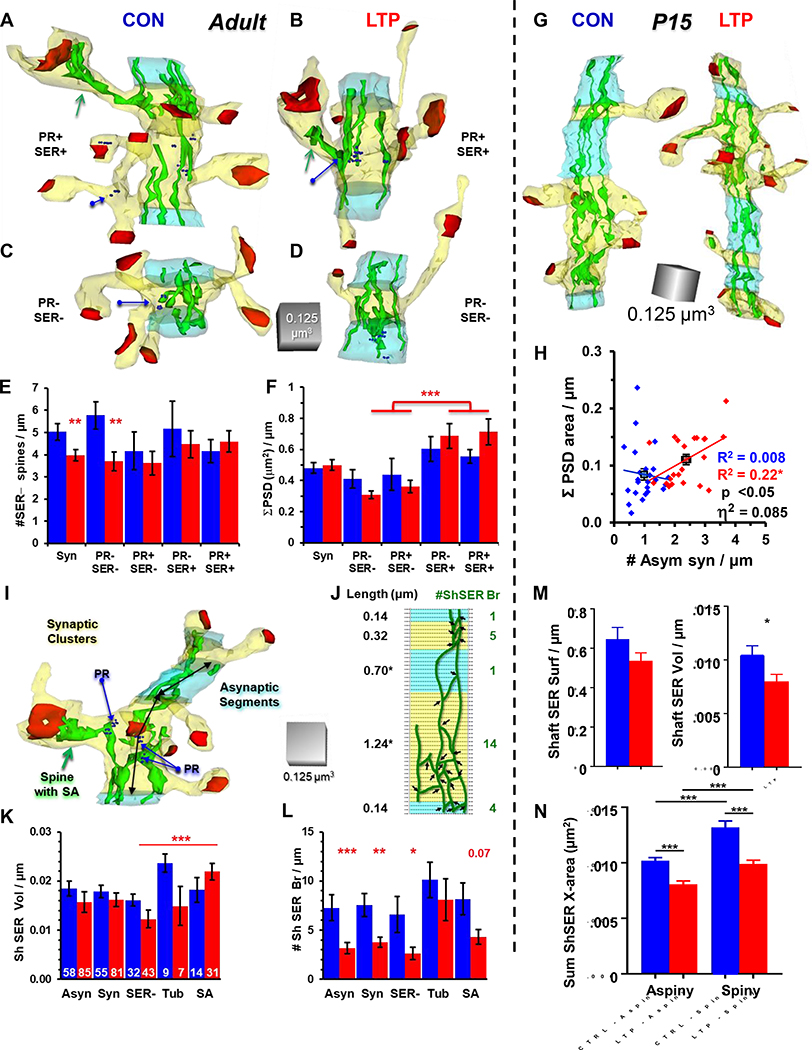
Nature teaches us that form precedes function, yet structure and function are intertwined. Such is the case with synapse structure, function, and plasticity underlying learning, especially in the hippocampus, a crucial brain region for memory formation. As the hippocampus matures, enduring changes in synapse structure produced by long-term potentiation (LTP) shift from synaptogenesis to synapse enlargement that is homeostatically balanced by stalled spine outgrowth and local spine clustering. Production of LTP leads to silent spine outgrowth at P15, and silent synapse enlargement in adult hippocampus at 2hours, but not at 5 or 30min following induction. Here we consider structural LTP in the context of developmental stage and variation in the availability of local resources of endosomes, smooth endoplasmic reticulum and polyribosomes. The emerging evidence supports a need for more nuanced analysis of synaptic plasticity in the context of subcellular resource availability and developmental stage.


Figures

Figure 1:
Figure 1:
LTP enhances synaptogenesis at P15 but stalls spine outgrowth in adults. A) 3DEMs of dendrites from oblique dendrites in s. radiatum of hippocampal area CA1 at P15 representing the 50th percentile rank. Spine density in the 2-hour (2h) control is less than (red <) the 2h LTP condition. B) Quantification of spine density (spines/ μm) length of dendrite in s. radiatum of perfusion-fixed hippocampus (PF), and after 5 minutes (5’), 30’, and 2 hr in the control or LTP conditions. C) 3DEMs of dendrites from adult hippocampal s. radiatum representing the 50th percentile rank. D) Plots show spine density in the 2h control is greater than (>) in the 2h LTP condition. Comparing PF between ages in B and D reveals natural age-dependent synaptogenesis. In all graphs, data were controlled for spine head diameter (HD). The LTP effects were not evident at 5 or 30 minutes; hence, these data are not plotted here, for simplicity, but they are available in the original publications. (Yellow – dendrite, green – smooth endoplasmic reticulum, red – excitatory postsynaptic density surface area. Gray asterisks indicate significant (p<0.05) differences between the indicated condition and PF; red asterisks show significant LTP effects. The medium blue asterisk in D, shows significant differences between time points for small spines. Adapted from Bourne and Harris, 2011; Bell et al., 2014; Watson et al., 2016; Kulik et al., 2019).
Figure 2:
Figure 2:
Role of SER and polyribosomes in supporting synaptic growth after LTP. A) Electron micrographs and A’) 3DEMs of dendritic spines without SER, with a simple tubule of SER, or a fully elaborated spine apparatus (SA). B) Following LTP in adults, the frequency of SER containing spines does not change; however, there is a significant shift from a single tubule (T) to the SA form of SER (*, p<0.5, n= number of spines in each condition). C) Electron micrograph and C’) 3DEM of spine containing a polyribosome, but no SER. D) The LTP-related synapse enlargement is minimal in spines lacking PR or SER, is greater on spines that retain PR, and is greatest on spines containing SER. Each graph illustrates the actual PSD areas, controlled for head diameter, and plotted on a log-normal scale, with correlation values (R2), and results of ANCOVA (p values and effect sizes, η2). E) At P15, most of the new spines produced 2 hours after LTP induction have small synapses and contain no SER. F) 3DEM of dendritic segment from P15 illustrating secretory compartments increase in spines after LTP. G) At P15, the increased secretory elements are primarily small vesicles (sv) or recycling compartments (RC) while some are also coated pits (cp) coated vesicles (cv) or large clear vesicles (LV). Amorphous vesicles and degradative structures are not elevated significantly.
Figure 3:
Figure 3:
Resource regulation of spine clusters. A-D) Representative dendritic segments from adult hippocampal slices under control and LTP conditions. Resource rich clusters have spines that contain PR or SER and resource poor clusters have no PR- or SER-containing spines. Each reconstruction is at about the 50th percentile rank within condition by spine density within the synaptic cluster (yellow) which is surrounded by an asynaptic region (light blue that is least 120 nm long, and averages 250 nm in both control and LTP conditions). E) The density of spines without SER is reduced overall in the synaptic clusters (Syn) following LTP, and this effect only occurred in clusters that lacked resource-rich spines (**p<0.01). F) Summed PSD area is balanced across all synaptic clusters and is greater following LTP in clusters that have resource-rich spines (***p<0.001). G) Representative dendritic segments from P15 hippocampal slices under control and LTP conditions. H) Synaptogenesis following LTP increases the mean (black squares) summed PSD area in proportion to the increase in spine density, with an overall effect size of 8% (η2). I-J) Calculating the number of SER branches per synaptic cluster or asynaptic segment length in adults. K) The volume of SER in the dendritic shaft increases following LTP in those clusters that had a spine with a spine apparatus (SA). L) Overall, the number of SER branches in the dendritic shaft decreases following LTP in adults but is retained in the clusters that contain spines with SER tubules (Tub) or SA. M) At P15, Shaft SER volume decreased as does the N) complexity of shaft SER in both aspiny and spiny segments. (Adapted from: Chirillo et al., 2019; Watson et al., 2015; and Kulik et al., 2019).
Figure 4:
Figure 4:
Model for silent synaptogenesis and synapse enlargement. A) Saturation of LTP and stable control responses. B) During STP at P15, GluAR are added to existing PSDs. By 120 min during LTP, new GluAR-lacking spines emerge (orange spines with light blue PSDs). C) In adults, some spines have GluAR-containing portions of the PSD, that are never-the-less silent because there are no presynaptic vesicles opposed to those zones (light blue zones in PSD, at 4 spines with presynaptic axonal boutons also illustrated; for simplicity, the other presynaptic axons are not illustrated at P15 or in adults). Zones of the PSD with presynaptic vesicles are red, being both pre- and postsynaptically active. In adults, the new spines that emerge during control stimulation lack GluARs. Induction of LTP blocks spine outgrowth (X’s) and fills presynaptic zones with vesicles (red arrow). By 120 minutes, new PSD areas are added that lack presynaptic active zones (blue arrow)

References

    1. Segal M, Dendritic spines: Morphological building blocks of memory. Neurobiol Learn Mem, 2017. 138: p. 3–9. - PubMed
    1. Nakahata Y and Yasuda R, Plasticity of Spine Structure: Local Signaling, Translation and Cytoskeletal Reorganization. Front Synaptic Neurosci, 2018. 10: p. 29. - PMC - PubMed
    1. Kruijssen DLH and Wierenga CJ, Single Synapse LTP: A Matter of Context? Front Cell Neurosci, 2019. 13: p. 496. - PMC - PubMed
    1. Bourne JN and Harris KM, Coordination of size and number of excitatory and inhibitory synapses results in a balanced structural plasticity along mature hippocampal CA1 dendrites during LTP. Hippocampus, 2011. 21(4): p. 354–373. - PMC - PubMed
    1. Watson DJ, et al., LTP enhances synaptogenesis in the developing hippocampus. Hippocampus, 2016. 26(5): p. 560–76. - PMC - PubMed

Publication types