. 2020 Jul 9;12(7):739.
doi: 10.3390/v12070739.

Infection of a Lepidopteran Cell Line with Deformed Wing Virus

Affiliations

Infection of a Lepidopteran Cell Line with Deformed Wing Virus

Tal Erez et al. Viruses. .

Abstract

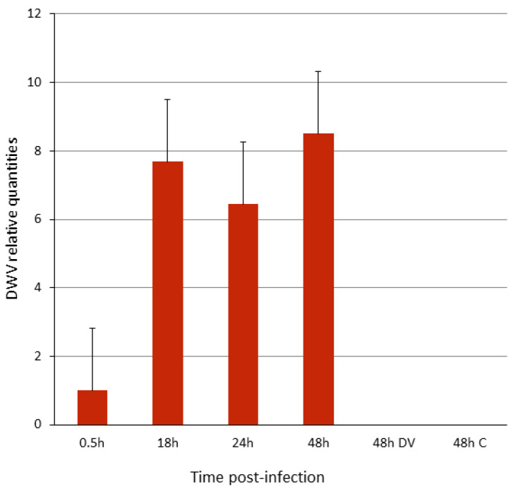
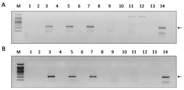
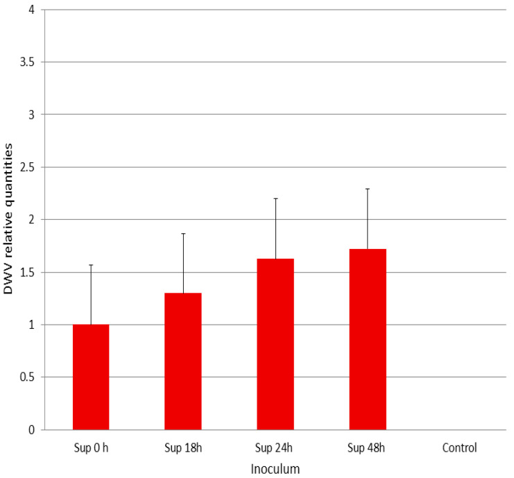
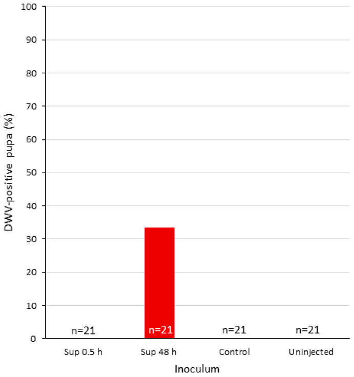
Many attempts to develop a reliable cell cultured-based system to study honey bee virus infections have encountered substantial difficulties. We investigated the ability of a cell line from a heterologous insect to sustain infection by a honey bee virus. For this purpose, we infected the Lepidopteran hemocytic cell line (P1) with Deformed wing virus (DWV). The genomic copies of DWV increased upon infection, as monitored by quantitative RT-PCR. Moreover, a tagged-primer-based RT-PCR analysis showed the presence of DWV negative-sense RNA in the cells, indicating virus replication. However, the DWV from infected cells was mildly infectious to P1 cells. Similar results were obtained when the virus was injected into Apis mellifera pupae. Thus, though the virus yields from the infected cells appeared to be very low, we show for the first time that DWV can replicate in a heterologous cell line. Given the availability of many other insect cell lines, our study paves the way for future exploration in this direction. In the absence of adequate A. mellifera cell lines, exploring the ability of alternative cell lines to enable honey bee virus infections could provide the means to study and understand the viral infectious cycle at the cellular level and facilitate obtaining purified isolates of these viruses.

Keywords: Deformed wing virus; Lepidopteran cells; honey bee virus.


Conflict of interest statement

The authors declare no conflict of interest. The funders had no role in the design of the study; in the collection, analysis, or interpretation of data; in the writing of the manuscript; or in the decision to publish the results.

Figures

Figure 1
Figure 1
Light micrograph of Deformed wing virus (DWV)-infected and mock-infected P1 cells. DWV-infected cells at 0, 24, and 48h post-infection (panels A, B, and C, respectively); D, mock-infected cells at 48 h. Bar 50 µm.
Figure 2
Figure 2
Infection of P1 cells with DWV. Y-axis, relative amount of DWV genomic copies, normalized expression in respect to the virus bound to the cells for 0.5 h of incubation and wash. X-axis, h, hours; DV, heat-inactivated virus inoculum; C, mock-infected cells (see Materials and Methods, Section 2.3). Bars represent standard error values. One-way ANOVA (F = 4.8581, P = 0.041).
Figure 3
Figure 3
Detection of the DWV negative-sense RNA strand in infected P1-cells. Panels A and B, PCR from cells incubated for 15 and 30 min with DWV and subsequent wash of the viral inoculum, respectively. Lanes 1 and 2—Samples extracted at 15 and 30 min p.i. (Panels A and B, respectively). Lanes 3–4, 5–6, 7–8—Samples extracted at 18, 24, and 48 h. p.i., respectively. Lanes 9–10—samples extracted from heat-inactivated DWV-infection at 48 h p.i. Lanes 11–12—Extracts from mock-infected cells. Even numbers indicate a PCR control reaction from the same individual RNA performed on the cDNA produced without any primer (see the section Materials and Methods, Section 2.4). M—100 bp DNA Ladder RTU Marker (GeneDireXTM), lower band 100 bp. Lane 13—NTC, non-template control, amplification of the positive sample without PCR primers. Lane 14, DWV amplicon (arrow).
Figure 4
Figure 4
Infectivity of DWV-P1 to P1 cells. Y-axis, relative amount of DWV genomic copies, normalized expression in respect to the virus bound to the cells for 0.5 h of incubation and wash (Sup. 0). X-axis, supernatant from P1 DWV-infected cells (DWV-P1) collected at various hours (h) p.i. used as inoculum. Control, supernatant from mock-infected cells used as inoculum. Bars represent standard error values. One-way ANOVA (F = 0.6954, P = 0.6333).
Figure 5
Figure 5
DWV prevalence of injected pupae. X-axis, individual pupae treated as follows: Injected with DWV-P1 at 0.5 and 48 h (Sup. 0.5 and 48 h, respectively) supernatant from mock-infected cells (control) or uninjected (see Materials and Methods, Section 2.5).

References

    1. Beaurepaire A., Doublet V., De Miranda J.R., Piot N., Antunez K., Campbell E., Chantawannakul P., Chejanovsky N., Gajda A., Heerman M., et al. Diversity and global distribution of viruses of the western honey bee, Apis mellifera. Insects. 2020;11:239. doi: 10.3390/insects11040239. - DOI - PMC - PubMed
    1. Chen Y.P., Siede R. Honey bee viruses. Adv. Virus Res. 2007;70:33–80. doi: 10.1016/S0065-3527(07)70002-7. - DOI - PubMed
    1. De Miranda J.R., Cordoni G., Budge G. The Acute bee paralysis virus—Kashmir bee virus—Israeli acute paralysis virus complex. J. Invertebr. Pathol. 2009;103(Suppl. 1):S30–S47. doi: 10.1016/j.jip.2009.06.014. - DOI - PubMed
    1. De Miranda J.R., Genersch E. Deformed wing virus. J. Invertebr. Pathol. 2010;103:S48–S61. doi: 10.1016/j.jip.2009.06.012. - DOI - PubMed
    1. De Miranda J.R., Dainat B., Locke B., Cordoni G., Berthoud H., Gauthier L., Neumann P., Budge G.E., Ball B.V., Stoltz D.B. Genetic characterization of slow bee paralysis virus of the honeybee (Apis mellifera L.) J. Gen. Virol. 2010;91:2524–2530. doi: 10.1099/vir.0.022434-0. - DOI - PubMed

Publication types

Supplementary concepts