

Correlation of MET and PD-L1 Expression in Malignant Melanoma
- PMID: 32659961
- PMCID: PMC7408820
- DOI: 10.3390/cancers12071847
Correlation of MET and PD-L1 Expression in Malignant Melanoma
Abstract
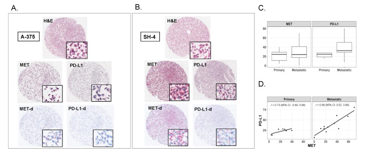
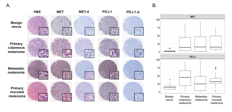
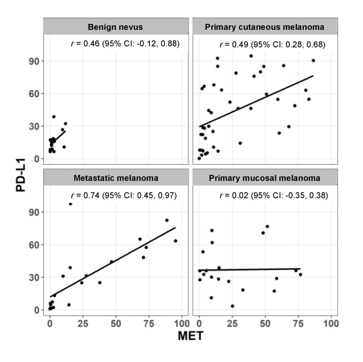
The proto-oncogene MET, the hepatocyte growth factor (HGF) receptor, is a transmembrane receptor tyrosine kinase (RTK) with a prominent role in tumor metastasis and resistance to anti-cancer therapies. Melanoma demonstrates relatively frequent MET aberrations, including MET gene amplification. Concurrently, programmed death-ligand 1 (PD-L1), with its ability to evade anti-tumor immune responses, has emerged as a prominent therapeutic target in melanoma and other malignancies and its expression is used as a predictive biomarker of response to immunotherapy. We performed immunohistochemistry analysis of MET and PD-L1 in 18 human melanoma cell lines derived from both primary and metastatic lesions, and in a human melanoma tissue microarray containing one hundreds melanocytic lesions, including primary cutaneous melanomas, primary mucosal melanomas, metastatic melanomas and benign melanocytic nevi as controls. After color deconvolution, each core was segmented to isolate staining and calculate the percentage of positive cells. Overall, MET expression was higher in tumors with increased PD-L1 expression. Moreover, a robust correlation between MET and PD-L1 expression was found in samples from metastatic melanoma and not in primary cutaneous or mucosal melanoma. These data suggest that relative expression levels of these proteins in combination is a marker of advanced disease and testing for expression of these markers should be considered in patients with melanoma.
Keywords: MET; PD-L1; melanoma; metastasis.
Conflict of interest statement
The authors declare no conflict of interest.
Figures
Similar articles
-
Comparative investigation of cell cycle and immunomodulatory genes in mucosal and cutaneous melanomas: Preliminary data suggest a potential promising clinical role for p16 and the PD-1/PD-L1 axis.Pathol Res Pract. 2022 Jan;229:153689. doi: 10.1016/j.prp.2021.153689. Epub 2021 Nov 22. Pathol Res Pract. 2022. PMID: 34844086
-
COX-2 expression positively correlates with PD-L1 expression in human melanoma cells.J Transl Med. 2017 Feb 23;15(1):46. doi: 10.1186/s12967-017-1150-7. J Transl Med. 2017. PMID: 28231855 Free PMC article.
-
Identification and clinical relevance of PD-L1 expression in primary mucosal malignant melanoma of the head and neck.Melanoma Res. 2015 Dec;25(6):503-9. doi: 10.1097/CMR.0000000000000197. Melanoma Res. 2015. PMID: 26352784
-
The Next Immune-Checkpoint Inhibitors: PD-1/PD-L1 Blockade in Melanoma.Clin Ther. 2015 Apr 1;37(4):764-82. doi: 10.1016/j.clinthera.2015.02.018. Epub 2015 Mar 29. Clin Ther. 2015. PMID: 25823918 Free PMC article. Review.
-
The Role of MET in Melanoma and Melanocytic Lesions.Am J Pathol. 2019 Nov;189(11):2138-2148. doi: 10.1016/j.ajpath.2019.08.002. Epub 2019 Aug 30. Am J Pathol. 2019. PMID: 31476283 Review.
Cited by
-
Multiomics integration reveals the effect of Orexin A on glioblastoma.Front Pharmacol. 2023 Jan 20;14:1096159. doi: 10.3389/fphar.2023.1096159. eCollection 2023. Front Pharmacol. 2023. PMID: 36744263 Free PMC article.
-
Exploration of Immune-Modulatory Effects of Amivantamab in Combination with Pembrolizumab in Lung and Head and Neck Squamous Cell Carcinoma.Cancer Res Commun. 2024 Jul 1;4(7):1748-1764. doi: 10.1158/2767-9764.CRC-24-0107. Cancer Res Commun. 2024. PMID: 38916448 Free PMC article.
-
LncRNA SNHG4 promotes malignant biological behaviors and immune escape of colorectal cancer cells by regulating the miR-144-3p/MET axis.Am J Transl Res. 2021 Oct 15;13(10):11144-11161. eCollection 2021. Am J Transl Res. 2021. PMID: 34786048 Free PMC article.
-
MET Oncogene Targeting for Cancer Immunotherapy.Int J Mol Sci. 2024 Jun 1;25(11):6109. doi: 10.3390/ijms25116109. Int J Mol Sci. 2024. PMID: 38892318 Free PMC article. Review.
-
Prognostic value of receptor tyrosine kinases in malignant melanoma patients: A systematic review and meta-analysis of immunohistochemistry.Front Oncol. 2022 Sep 23;12:819051. doi: 10.3389/fonc.2022.819051. eCollection 2022. Front Oncol. 2022. PMID: 36212475 Free PMC article.
References
Grants and funding
LinkOut - more resources
Full Text Sources
Research Materials
Miscellaneous
