

The Role of Dysfunctional Adipose Tissue in Pancreatic Cancer: A Molecular Perspective
- PMID: 32659999
- PMCID: PMC7408631
- DOI: 10.3390/cancers12071849
The Role of Dysfunctional Adipose Tissue in Pancreatic Cancer: A Molecular Perspective
Abstract
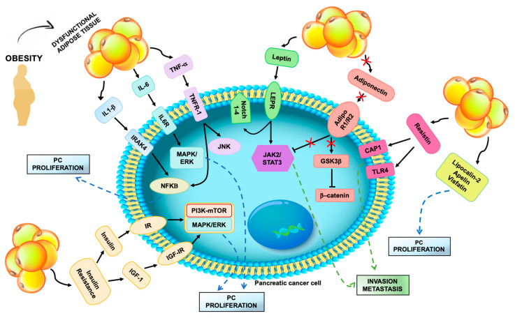
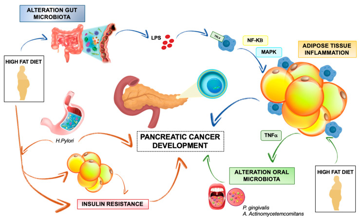
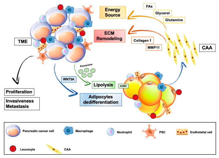
Pancreatic cancer (PC) is a lethal malignancy with rising incidence and limited therapeutic options. Obesity is a well-established risk factor for PC development. Moreover, it negatively affects outcome in PC patients. Excessive fat accumulation in obese, over- and normal-weight individuals induces metabolic and inflammatory changes of adipose tissue microenvironment leading to a dysfunctional adipose "organ". This may drive the association between abnormal fat accumulation and pancreatic cancer. In this review, we describe several molecular mechanisms that underpin this association at both local and systemic levels. We focus on the role of adipose tissue-derived circulating factors including adipokines, hormones and pro-inflammatory cytokines, as well as on the impact of the local adipose tissue in promoting PC. A discussion on potential therapeutic interventions, interfering with pro-tumorigenic effects of dysfunctional adipose tissue in PC, is included. Considering the raise of global obesity, research efforts to uncover the molecular basis of the relationship between pancreatic cancer and adipose tissue dysfunction may provide novel insights for the prevention of this deadly disease. In addition, these efforts may uncover novel targets for personalized interventional strategies aimed at improving the currently unsatisfactory PC therapeutic options.
Keywords: adipose tissue; obesity; pancreatic cancer.
Conflict of interest statement
The authors declare no conflict of interest.
Figures
Similar articles
-
Crosstalk between pancreatic cancer and adipose tissue: Molecular mechanisms and therapeutic implications.Biochem Biophys Res Commun. 2024 Dec 25;740:151012. doi: 10.1016/j.bbrc.2024.151012. Epub 2024 Nov 15. Biochem Biophys Res Commun. 2024. PMID: 39561650 Review.
-
Obesity and pancreatic cancer: An update of epidemiological evidence and molecular mechanisms.Pancreatology. 2019 Oct;19(7):941-950. doi: 10.1016/j.pan.2019.08.008. Epub 2019 Aug 16. Pancreatology. 2019. PMID: 31447281 Review.
-
New insights into the role of adipocytes in pancreatic cancer progression: paving the way towards novel therapeutic targets.Theranostics. 2023 Jul 3;13(12):3925-3942. doi: 10.7150/thno.82911. eCollection 2023. Theranostics. 2023. PMID: 37554282 Free PMC article. Review.
-
Obesity and Pancreatic Cancer: Insight into Mechanisms.Cancers (Basel). 2021 Oct 10;13(20):5067. doi: 10.3390/cancers13205067. Cancers (Basel). 2021. PMID: 34680216 Free PMC article. Review.
-
Inflammatory processes in obesity: focus on endothelial dysfunction and the role of adipokines as inflammatory mediators.Int Rev Immunol. 2019;38(4):157-171. doi: 10.1080/08830185.2019.1638921. Epub 2019 Jul 9. Int Rev Immunol. 2019. PMID: 31286783 Review.
Cited by
-
Obesity and Overweight Are Associated with Minimal Extrathyroidal Extension, Multifocality and Bilaterality of Papillary Thyroid Cancer.J Clin Med. 2021 Mar 2;10(5):970. doi: 10.3390/jcm10050970. J Clin Med. 2021. PMID: 33801171 Free PMC article.
-
Incorporating adipose tissue into a CT-based deep learning nomogram to differentiate granulomas from lung adenocarcinomas.iScience. 2024 Aug 19;27(10):110733. doi: 10.1016/j.isci.2024.110733. eCollection 2024 Oct 18. iScience. 2024. PMID: 39474083 Free PMC article.
-
Modifiable and Non-Modifiable Risk Factors for the Development of Non-Hereditary Pancreatic Cancer.Medicina (Kaunas). 2022 Jul 22;58(8):978. doi: 10.3390/medicina58080978. Medicina (Kaunas). 2022. PMID: 35893093 Free PMC article. Review.
-
Studies on Treatment Within the Scope of Medical Biotechnology for Pancreatic Diseases.Mol Biotechnol. 2025 Apr;67(4):1321-1335. doi: 10.1007/s12033-024-01142-5. Epub 2024 Apr 16. Mol Biotechnol. 2025. PMID: 38627328 Review.
-
The Role of Local Angiotensin II/Angiotensin Type 1-receptor Mechanisms in Adipose Tissue Dysfunction to Promote Pancreatic Cancer.Curr Cancer Drug Targets. 2024;24(12):1187-1194. doi: 10.2174/0115680096281059240103154836. Curr Cancer Drug Targets. 2024. PMID: 38347780 Review.
References
-
- Ferlay J.E.M., Lam F., Colombet M., Mery L., Pineros M., Znaor A. SI Global Cancer Observatory: Cancer Today. International Agency for Research on Cancer; Lyon, France: [(accessed on 30 March 2020)]. Available online: https//gco.iarc.fr/today.
Publication types
LinkOut - more resources
Full Text Sources
