Review
. 2020 Jul 9;9(7):1652.
doi: 10.3390/cells9071652.

COVID-19, Renin-Angiotensin System and Endothelial Dysfunction

Affiliations
Review

COVID-19, Renin-Angiotensin System and Endothelial Dysfunction

Razie Amraei et al. Cells. .

Abstract

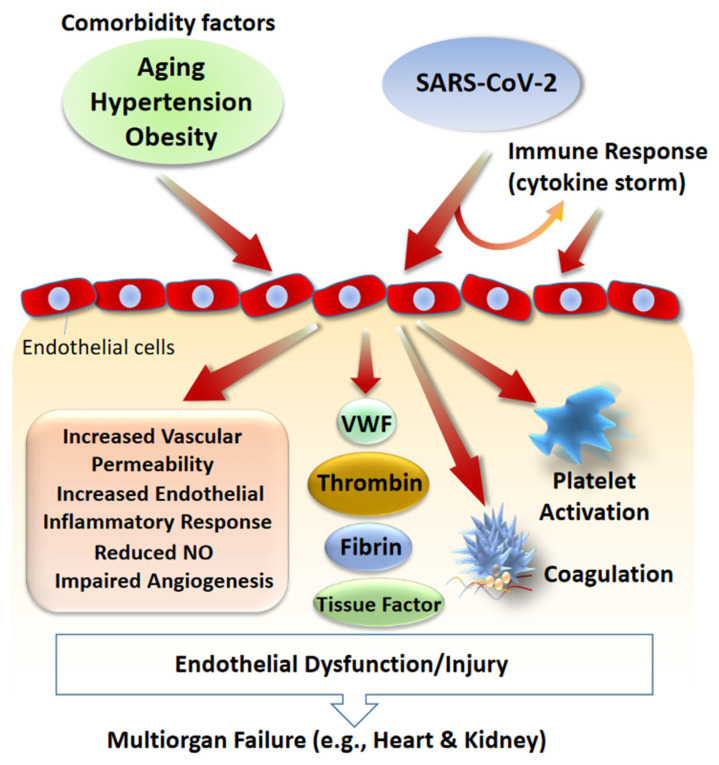
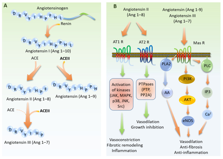
The newly emergent novel coronavirus disease 2019 (COVID-19) outbreak, which is caused by SARS-CoV-2 virus, has posed a serious threat to global public health and caused worldwide social and economic breakdown. Angiotensin-converting enzyme 2 (ACE2) is expressed in human vascular endothelium, respiratory epithelium, and other cell types, and is thought to be a primary mechanism of SARS-CoV-2 entry and infection. In physiological condition, ACE2 via its carboxypeptidase activity generates angiotensin fragments (Ang 1-9 and Ang 1-7), and plays an essential role in the renin-angiotensin system (RAS), which is a critical regulator of cardiovascular homeostasis. SARS-CoV-2 via its surface spike glycoprotein interacts with ACE2 and invades the host cells. Once inside the host cells, SARS-CoV-2 induces acute respiratory distress syndrome (ARDS), stimulates immune response (i.e., cytokine storm) and vascular damage. SARS-CoV-2 induced endothelial cell injury could exacerbate endothelial dysfunction, which is a hallmark of aging, hypertension, and obesity, leading to further complications. The pathophysiology of endothelial dysfunction and injury offers insights into COVID-19 associated mortality. Here we reviewed the molecular basis of SARS-CoV-2 infection, the roles of ACE2, RAS signaling, and a possible link between the pre-existing endothelial dysfunction and SARS-CoV-2 induced endothelial injury in COVID-19 associated mortality. We also surveyed the roles of cell adhesion molecules (CAMs), including CD209L/L-SIGN and CD209/DC-SIGN in SARS-CoV-2 infection and other related viruses. Understanding the molecular mechanisms of infection, the vascular damage caused by SARS-CoV-2 and pathways involved in the regulation of endothelial dysfunction could lead to new therapeutic strategies against COVID-19.

Keywords: ACE2; CD209L; L-SIGN; SARS-CoV-2; endothelial cell injury; endothelial dysfunction.


Conflict of interest statement

Authors declare no conflict of interest.

Figures

Figure 1
Figure 1
Role of comorbidity factors and SARS-CoV-2 in vascular dysfunction and vascular injury. Endothelial dysfunction is associated with aging and conditions such as hypertension and diabetes. SARS-CoV-2 can induce vascular damage directly or indirectly by stimulating immune response which results in excessive cytokine production (cytokine storm) which also can lead to vascular damage. SARS-CoV-2 induced vascular damage alone or in combination with pre-existing endothelial dysfunction can lead to multisystem organ failure and death. Key biochemical factors and cellular responses involved in the SARS-CoV-2 induced endothelial damage and endothelial dysfunction are shown.
Figure 2
Figure 2
Schematic and domain structure of angiotensin-converting enzyme 2 (ACE2). (A) General domain information including, ion binding, proteolytic cleavage sites and S protein binding motif are shown. (B) Crystal structure of ACE2 and location of ion bindings and catalytic domain in complex with ACE2 inhibitor, MLN-476, is shown.
Figure 3
Figure 3
The physiological role angiotensin system in cardiovascular system. (A) Angiotensinogen is cleaved by renin and produces angiotensin I (Ang 1–10). Ang 1–10 is substrate for both ACE and ACE2. While ACE generates Ang 1–8, ACE2 cleaves Ang-10 and generates Ang 1–9. Ang 1–8 is a major substrate for ACE2 which produces Ang 1–7. (B) Ang (1–8) serves as a ligand for G-protein-coupled receptors (GPCRs), AT1R and AT2R. Activation of AT1R promotes vasoconstriction, fibrotic remodeling, and inflammation. Stimulation of AT2R leads to vasodilation and growth inhibition. On the other hand, peptides produced by ACE2 (Ang 1–9 and Ang 1–7) bind to GPCR, Mas receptor leading to its activation and promotes vasodilation, anti-fibrosis, and anti-inflammation effects. AA, Arachidonic acid.
Figure 4
Figure 4
Cell adhesion of molecules are common receptors for viruses. Schematic of the most common cell adhesion molecules involved in virus recognition and viral cell entry. Coxsackievirus-adenovirus receptor (CAR) [127], intercellular adhesion molecule 1 (ICAM 1/CD54) [128], junctional adhesion molecule A (JAM-A) [129,130], and poliovirus receptor (PVR/CD155) [131]. MERS-CoV also employs carcinoembryonic antigen-related cell adhesion molecule 5 (CEACAM5) [132]. CLEC4M, (also known as L-SIGN and CD209L) and CAM, dendritic cell-specific ICAM-3 grabbing non-integrin (DC-SIGN, also called CD209) binds to Ebolavirus [133], Hepatitis C virus [134], human coronavirus 229E [135], Human cytomegalovirus/HHV-5 [136], Influenza virus [137], West-Nile virus [136], Japanese encephalitis virus [138], SARS-CoV [136,139,140], and SARS-CoV-2 [120].

References

    1. Wu Z., McGoogan J.M. Characteristics of and Important Lessons from the Coronavirus Disease 2019 (COVID-19) Outbreak in China: Summary of a Report of 72 314 Cases From the Chinese Center for Disease Control and Prevention. JAMA. 2020;323:1239–1242. doi: 10.1001/jama.2020.2648. - DOI - PubMed
    1. Lee N., Hui D., Wu A., Chan P., Cameron P., Joynt G.M., Ahuja A., Yung M.Y., Leung C.B., To K.F., et al. A major outbreak of severe acute respiratory syndrome in Hong Kong. N. Engl. J. Med. 2003;348:1986–1994. doi: 10.1056/NEJMoa030685. - DOI - PubMed
    1. Yu I.T., Li Y., Wong T.W., Tam W., Chan A.T., Lee J.H., Leung D.Y., Ho T. Evidence of airborne transmission of the severe acute respiratory syndrome virus. N. Engl. J. Med. 2004;350:1731–1739. doi: 10.1056/NEJMoa032867. - DOI - PubMed
    1. Huang C., Wang Y., Li X., Ren L., Zhao J., Hu Y., Zhang L., Fan G., Xu J., Gu X., et al. Clinical features of patients infected with 2019 novel coronavirus in Wuhan, China. Lancet. 2020;395:497–506. doi: 10.1016/S0140-6736(20)30183-5. - DOI - PMC - PubMed
    1. Ren L.L., Wang Y.M., Wu Z.Q., Xiang Z.C., Guo L., Xu T., Jiang Y.Z., Xiong Y., Li Y.J., Li X.W., et al. Identification of a novel coronavirus causing severe pneumonia in human: A descriptive study. Chin. Med. J. (Engl.) 2020 doi: 10.1097/CM9.0000000000000722. - DOI - PMC - PubMed

Publication types

MeSH terms

LinkOut - more resources