. 2020 Jul 13;20(1):283.
doi: 10.1186/s12886-020-01540-8.

Choroidal origin of endogenous Candida endophthalmitis

Affiliations

Choroidal origin of endogenous Candida endophthalmitis

Mark P Breazzano. BMC Ophthalmol. .

Abstract

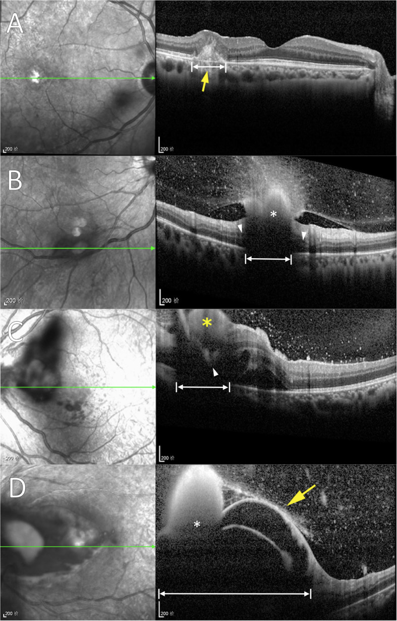
Endogenous Candida endophthalmitis (ECE) has been established with microscopic histopathology, both by autopsy and experimentation, to primarily originate from and involve the choroid. Zhuang et al. examined a series of patients with ECE using spectral-domain optical coherence tomography (SD-OCT) imaging and present a new classification scheme. The authors conclude the majority of lesions are primarily retinal in location without report of choroidal involvement. This discrepancy may be explained by posterior shadowing artifact and lack of discernment from associated retinal findings like infarction. These considerations are necessary in reviewing SD-OCT, characterizing ECE, and proposing new classification systems.

Keywords: Artifact; Choroid; Endogenous Candida endophthalmitis; Spectral-domain optical coherence tomography.


Conflict of interest statement

None.

Figures

Fig. 1
Fig. 1
Imaging of endogenous Candida endophthalmitis (ECE) demonstrated by Zhuang et al. [1], now with labeling overlay (white markings). These examples of ECE (classified as “retinal” lesion types 1–4, respectively) demonstrate primary choroidal involvement (a), and shadowing that cannot establish a retinal origin or exclude choroidal origin (b-d). a Disrupted architecture of inner choroid (double arrow) extending into the overlying retinal pigment epithelium and neurosensory retina is consistent with a primarily choroidal infiltrative and inflammatory process. b Posterior shadowing (double arrow) is found approximate in size to the overlying, anterior aspect of a large hyperreflective structure (*). Lesion origin cannot be determined because the view of the choroid and outer retina is overshadowed (double arrows), but cystic changes are apparent in outer retina adjacent to the edges of shadowing (arrowheads) which suggest a possibly deep lesion origin. c Posterior extent of hyperreflectivity is seen extending at least into deep retina (arrowhead), surrounded by neurosensory retinal detachment and extensive shadowing that completely obscures underlying choroid (double arrows). d Large hyperreflective mass (*) with florid inner retinoschisis and secondary shadowing (double arrows) blocks choroidal visualization

References

    1. Zhuang H, Ding X, Gao F, Zhang T, Ni Y, Chang Q, Xu G. Optical coherence tomography features of retinal lesions in Chinese patients with endogenous Candida endophthalmitis. BMC Ophthalmol. 2020;20(1):52. doi: 10.1186/s12886-020-01337-9. - DOI - PMC - PubMed
    1. Griffin JR, Pettit TH, Fishman LS, Foos RY. Blood-borne Candida endophthalmitis. A clinical and pathologic study of 21 cases. Arch Ophthalmol. 1973;89(6):450–460. doi: 10.1001/archopht.1973.01000040452002. - DOI - PubMed
    1. Edwards JE, Jr, Foos RY, Montgomerie JZ, Guze LB. Ocular manifestations of Candida septicemia: review of seventy-six cases of hematogenous Candida endophthalmitis. Medicine (Baltimore) 1974;53(1):47–75. doi: 10.1097/00005792-197401000-00002. - DOI - PubMed
    1. McDonnell PJ, McDonnell JM, Brown RH, Green WR. Ocular involvement in patients with fungal infections. Ophthalmology. 1985;92(5):706–709. doi: 10.1016/S0161-6420(85)33989-1. - DOI - PubMed
    1. Edwards JE, Jr, Montgomerie JZ, Foos RY, Shaw VK, Guze LB. Experimental hematogenous endophthalmitis caused by Candida albicans. J Infect Dis. 1975;131(6):649–657. doi: 10.1093/infdis/131.6.649. - DOI - PubMed

LinkOut - more resources