

Structural basis of a shared antibody response to SARS-CoV-2
- PMID: 32661058
- PMCID: PMC7402627
- DOI: 10.1126/science.abd2321
Structural basis of a shared antibody response to SARS-CoV-2
Abstract
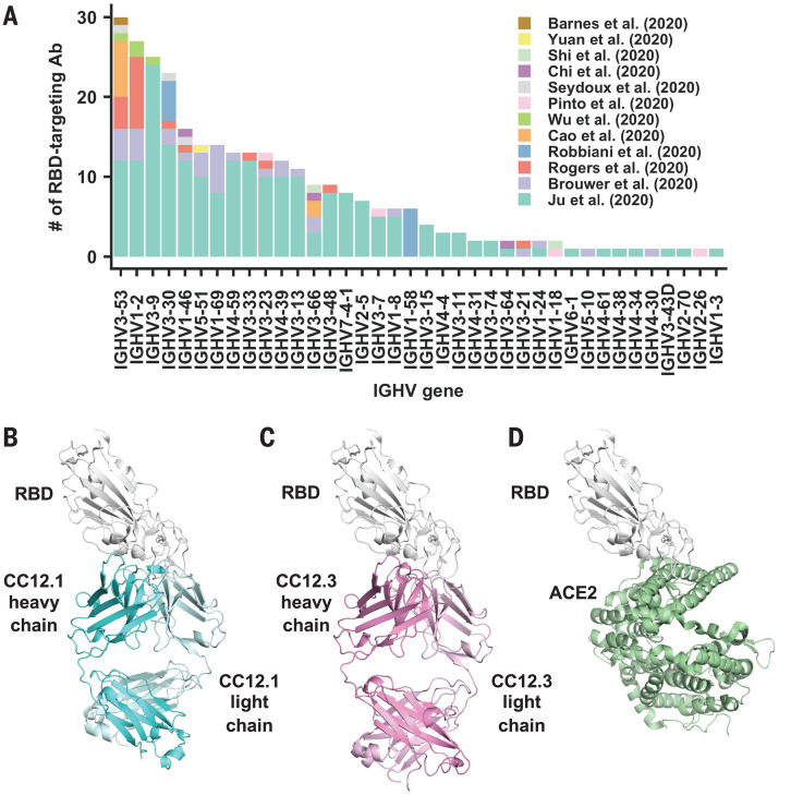
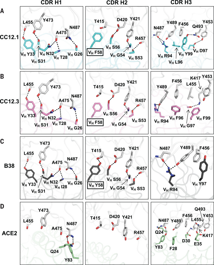
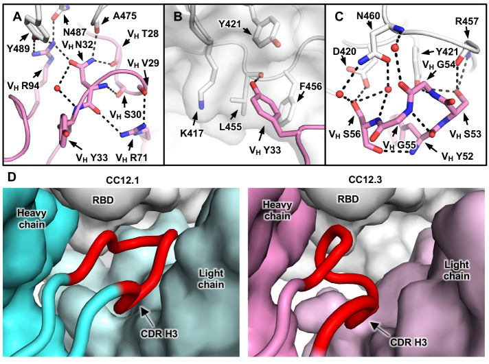
Molecular understanding of neutralizing antibody responses to severe acute respiratory syndrome coronavirus 2 (SARS-CoV-2) could accelerate vaccine design and drug discovery. We analyzed 294 anti-SARS-CoV-2 antibodies and found that immunoglobulin G heavy-chain variable region 3-53 (IGHV3-53) is the most frequently used IGHV gene for targeting the receptor-binding domain (RBD) of the spike protein. Co-crystal structures of two IGHV3-53-neutralizing antibodies with RBD, with or without Fab CR3022, at 2.33- to 3.20-angstrom resolution revealed that the germline-encoded residues dominate recognition of the angiotensin I converting enzyme 2 (ACE2)-binding site. This binding mode limits the IGHV3-53 antibodies to short complementarity-determining region H3 loops but accommodates light-chain diversity. These IGHV3-53 antibodies show minimal affinity maturation and high potency, which is promising for vaccine design. Knowledge of these structural motifs and binding mode should facilitate the design of antigens that elicit this type of neutralizing response.
Copyright © 2020 The Authors, some rights reserved; exclusive licensee American Association for the Advancement of Science. No claim to original U.S. Government Works.
Figures

Update of
-
Structural basis of a public antibody response to SARS-CoV-2.bioRxiv [Preprint]. 2020 Jun 9:2020.06.08.141267. doi: 10.1101/2020.06.08.141267. bioRxiv. 2020. Update in: Science. 2020 Aug 28;369(6507):1119-1123. doi: 10.1126/science.abd2321. PMID: 32577642 Free PMC article. Updated. Preprint.
Comment in
-
Molecular features of IGHV3-53-encoded antibodies elicited by SARS-CoV-2.Signal Transduct Target Ther. 2020 Aug 25;5(1):170. doi: 10.1038/s41392-020-00287-4. Signal Transduct Target Ther. 2020. PMID: 32843617 Free PMC article. No abstract available.
References
-
- Parameswaran P., Liu Y., Roskin K. M., Jackson K. K. L., Dixit V. P., Lee J.-Y., Artiles K. L., Zompi S., Vargas M. J., Simen B. B., Hanczaruk B., McGowan K. R., Tariq M. A., Pourmand N., Koller D., Balmaseda A., Boyd S. D., Harris E., Fire A. Z., Convergent antibody signatures in human dengue. Cell Host Microbe 13, 691–700 (2013). 10.1016/j.chom.2013.05.008 - DOI - PMC - PubMed
-
- Pieper K., Tan J., Piccoli L., Foglierini M., Barbieri S., Chen Y., Silacci-Fregni C., Wolf T., Jarrossay D., Anderle M., Abdi A., Ndungu F. M., Doumbo O. K., Traore B., Tran T. M., Jongo S., Zenklusen I., Crompton P. D., Daubenberger C., Bull P. C., Sallusto F., Lanzavecchia A., Public antibodies to malaria antigens generated by two LAIR1 insertion modalities. Nature 548, 597–601 (2017). 10.1038/nature23670 - DOI - PMC - PubMed
Publication types
MeSH terms
Substances
Grants and funding
LinkOut - more resources
Full Text Sources
Other Literature Sources
Miscellaneous
