Review
. 2020 Sep 11;295(37):12910-12934.
doi: 10.1074/jbc.REV120.013930. Epub 2020 Jul 13.

The molecular virology of coronaviruses

Affiliations
Review

The molecular virology of coronaviruses

Ella Hartenian et al. J Biol Chem. .

Abstract

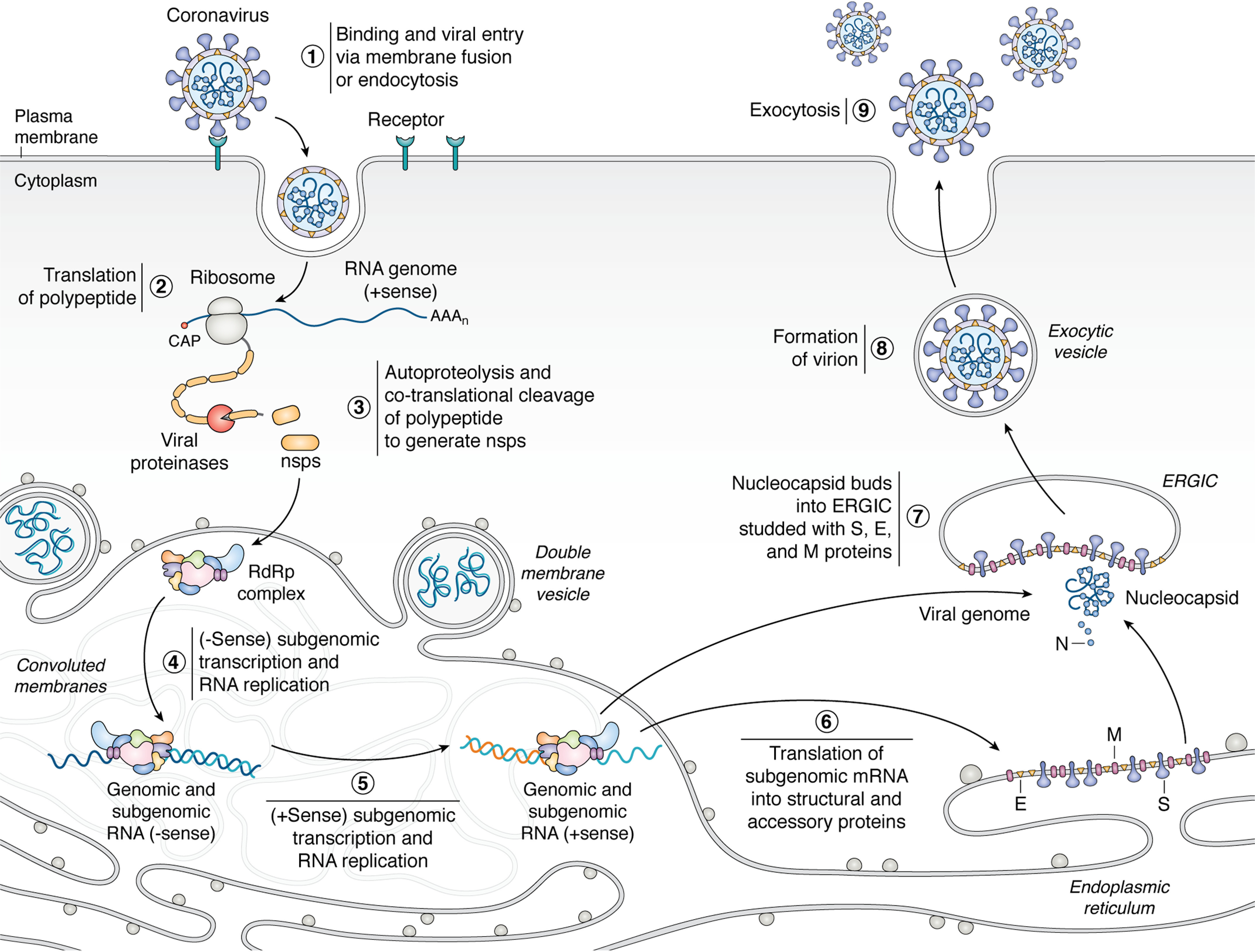
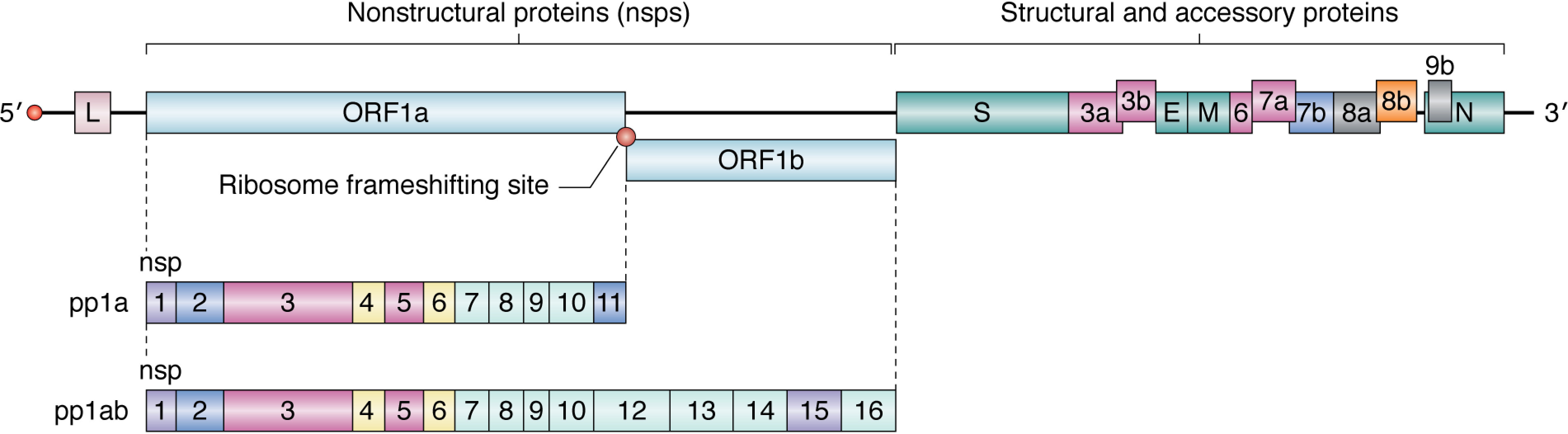
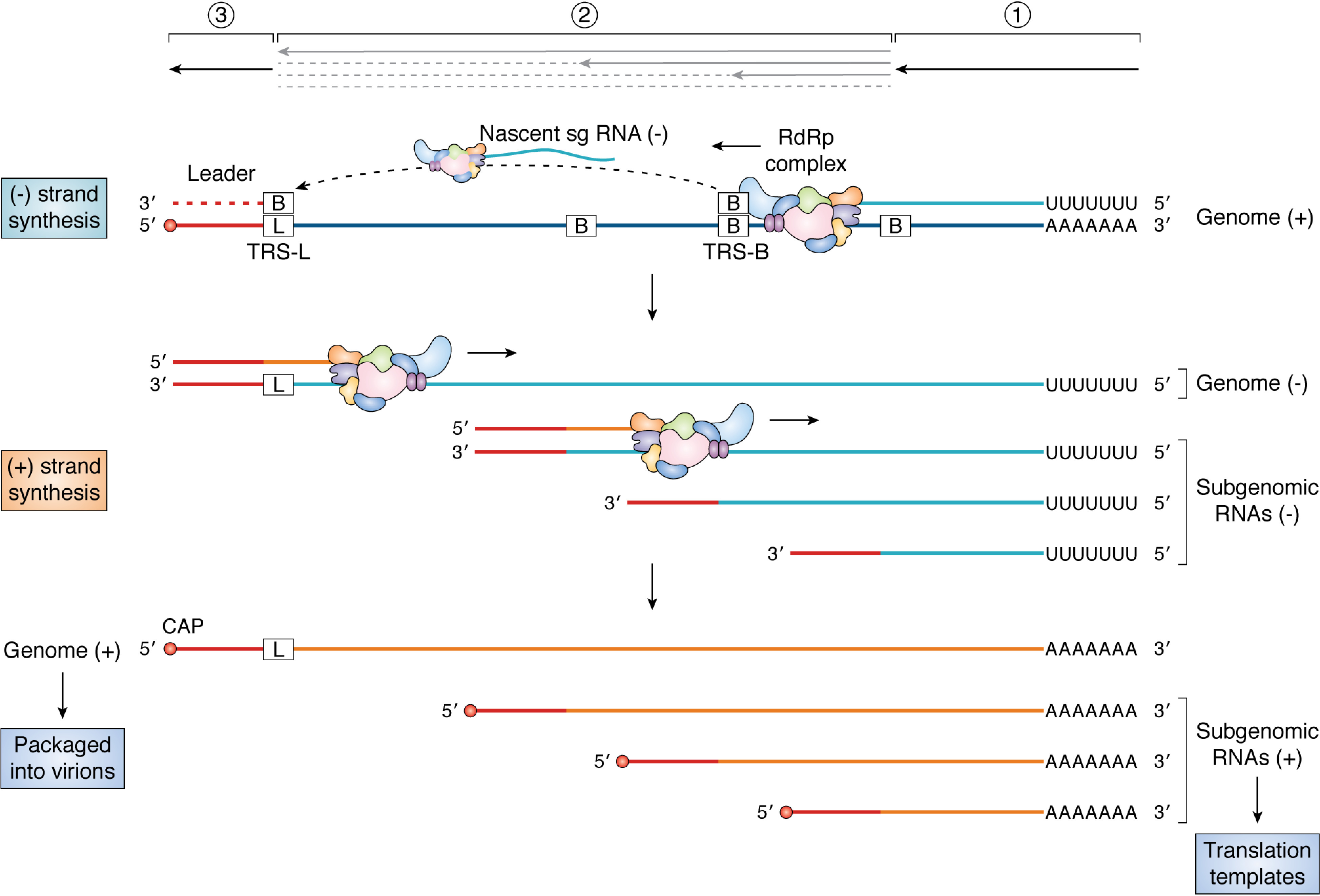
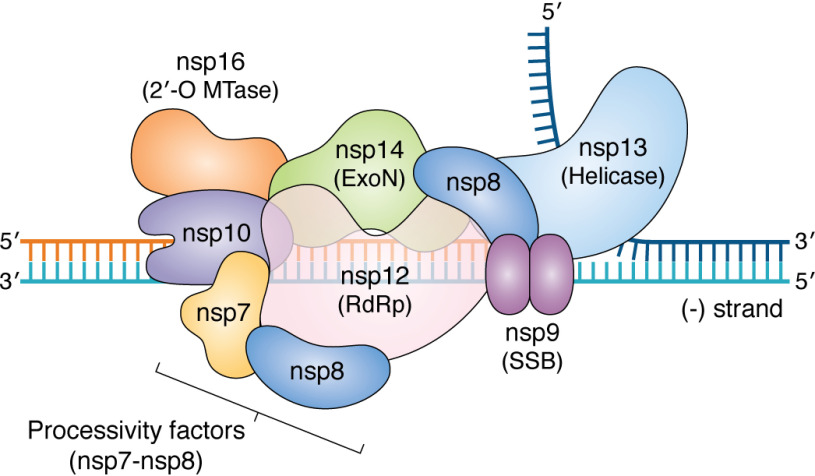
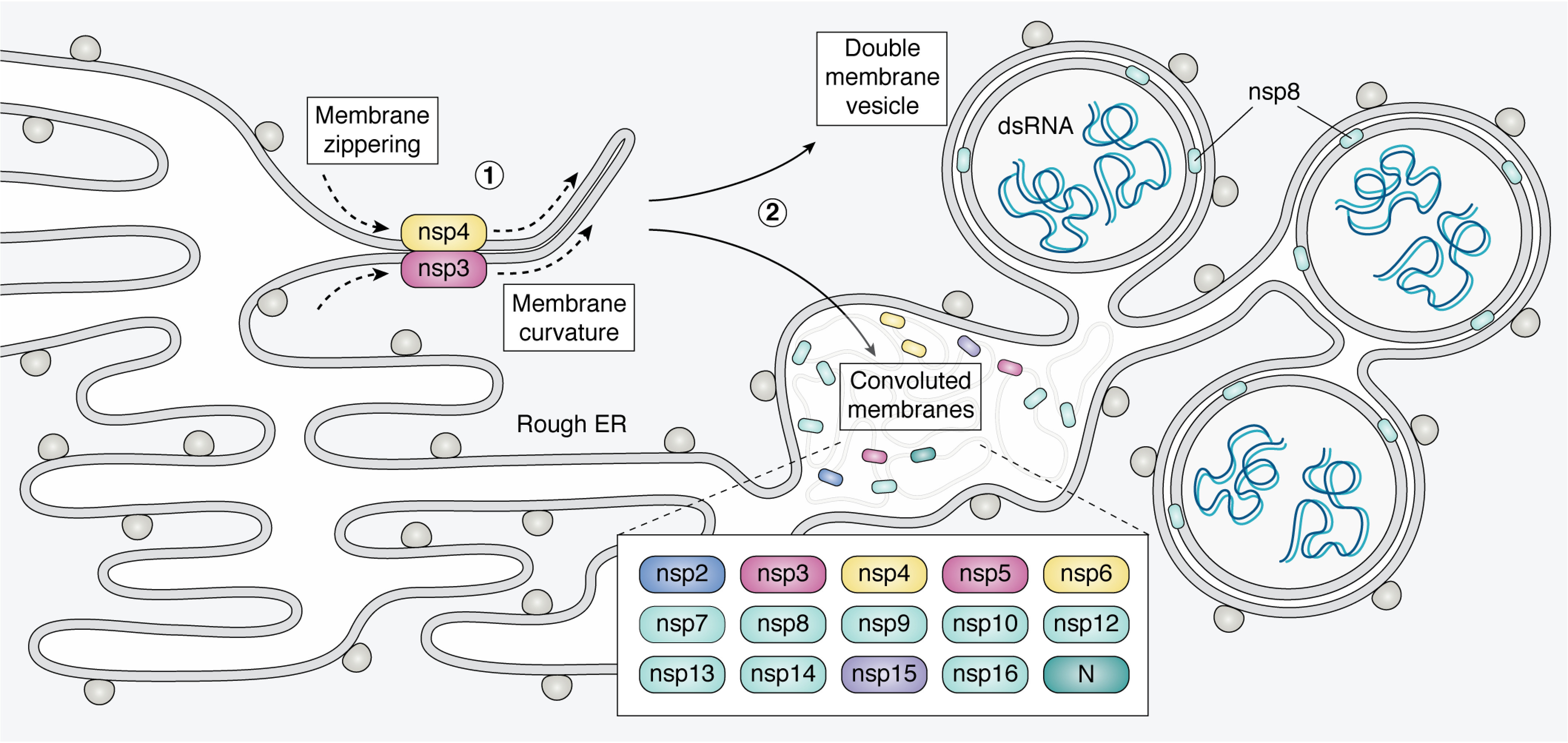
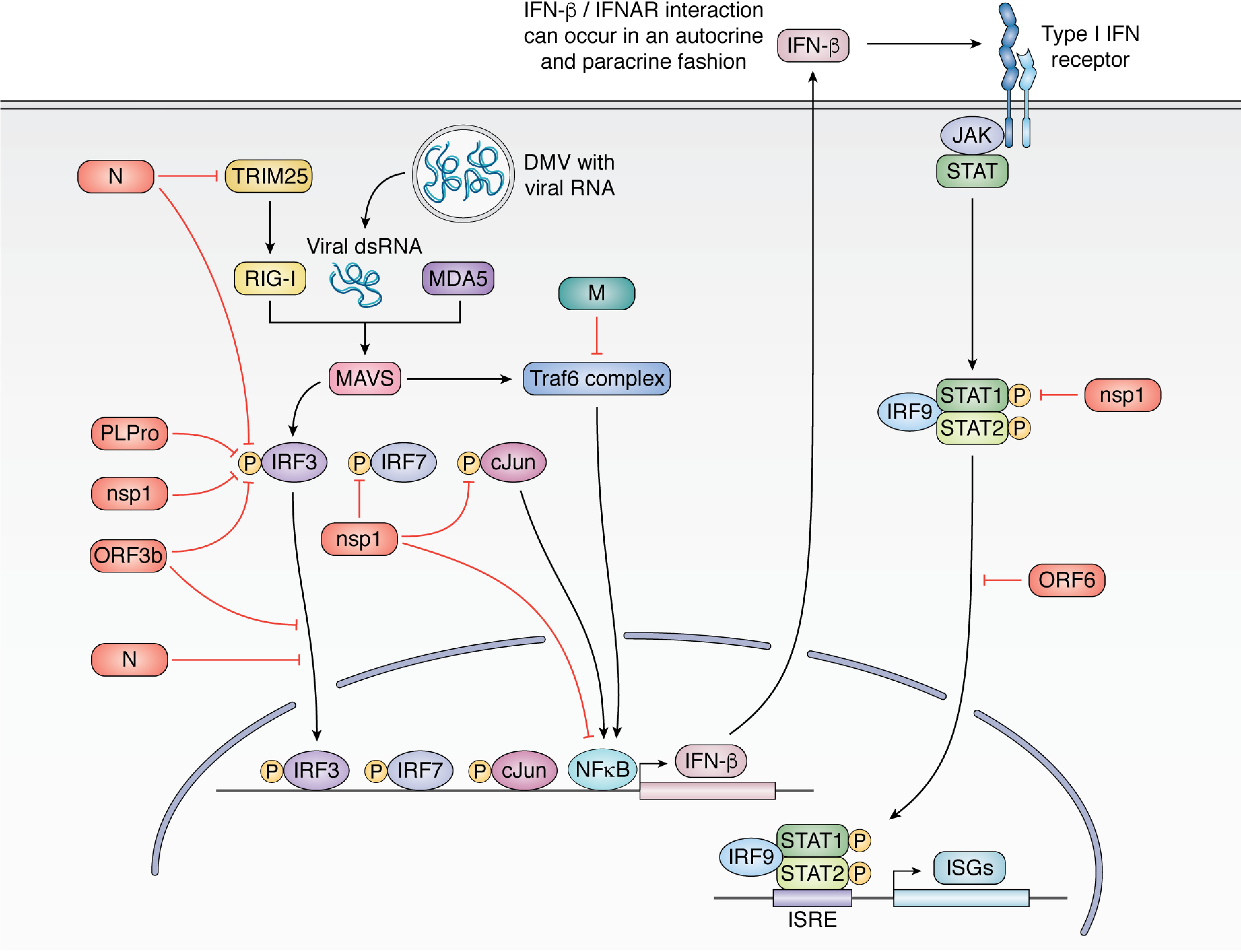
Few human pathogens have been the focus of as much concentrated worldwide attention as severe acute respiratory syndrome coronavirus 2 (SARS-CoV-2), the cause of COVID-19. Its emergence into the human population and ensuing pandemic came on the heels of severe acute respiratory syndrome coronavirus (SARS-CoV) and Middle East respiratory syndrome coronavirus (MERS-CoV), two other highly pathogenic coronavirus spillovers, which collectively have reshaped our view of a virus family previously associated primarily with the common cold. It has placed intense pressure on the collective scientific community to develop therapeutics and vaccines, whose engineering relies on a detailed understanding of coronavirus biology. Here, we present the molecular virology of coronavirus infection, including its entry into cells, its remarkably sophisticated gene expression and replication mechanisms, its extensive remodeling of the intracellular environment, and its multifaceted immune evasion strategies. We highlight aspects of the viral life cycle that may be amenable to antiviral targeting as well as key features of its biology that await discovery.

Keywords: RNA polymerase; SARS-CoV-2; cellular immune response; coronavirus; endoplasmic reticulum; endoplasmic reticulum (ER); innate immunity; pathogenesis; plus-stranded RNA virus; viral replication; virology; virus; virus entry.


Conflict of interest statement

Conflict of interest—The authors declare that they have no conflicts of interest with the contents of this article.

Figures

Figure 1.
Figure 1.
Coronaviruses engage with a host cell-surface receptor and deposit their RNA genomes into the host cytoplasm through endocytosis or directmembrane fusion (1). The positive-sense RNA genome is translated by the host translation machinery (2) to make polyproteins that are cotranslationally cleaved by proteases encoded in the polyprotein to generate components of RdRp complex (3). The RdRp complex uses the genome as a template to generate negative-sense subgenome and genome-length RNAs (4), which are in turn used as templates for synthesis of positive-sense full-length progeny genomes and subgenomic mRNAs (5). Transcription and replication occur in convoluted membranes (CM) adjacent to DMVs that are both derived from rough endoplasmic reticulum(see Fig. 6 for more details). The subgenomic mRNAs are translated into structural and accessory proteins (6). The positive sense genomic RNA is bound by nucleocapsid and buds into the ERGIC, which is decorated with structural proteins S, E, and M translated from positive-sense subgenomic RNAs (steps 6 and 7). The enveloped virion is then exported from the cell by exocytosis (steps 8 and 9).
Figure 2.
Figure 2.
Mechanism of SARS-CoV-2 viral entry. The SARS-CoV-2 S protein engages with the host ACE2 receptor and is subsequently cleaved at S1/S2 and S2′ sites by TMPRSS2 protease. This leads to activation of the S2 domain and drives fusion of the viral and host membranes. See section on 'viral entry' for details.
Figure 3.
Figure 3.
Genome organization of SARS-CoV. The RNA genome encodes two categories of proteins: nsps and structural and accessory proteins. The nonstructural proteins are encoded in ORF1a and ORF1b. Cap-dependent translation begins at ORF1a and produces pp1a, encompassing nsp1–11, or pp1ab, a longer polypeptide that includes nsp12–16. The production of either polypeptide depends on whether the stop codon at ORF1a is recognized by the ribosome or is bypassed through a change in the reading frame by the ribosome frameshifting site. The structural and accessory proteins are synthesized by translation of their respective subgenomic mRNAs (see Fig. 4). The proteins have been color-coded by functional categories for SARS-CoV (see Table 1).
Figure 4.
Figure 4.
Discontinuous transcription. The RdRp complex initiates transcription at the 39 end of the positive-sense genome (1). Upon copying the TRS-B sequences present at specific sites along the genome body (2), the RdRp complex may “jump” to the TRS-L sequence (3) owing to complementarity between the TRS-B sequence on the nascent sg RNA and TRS-L sequence on the genome. Transcription is resumed on the new template, and the leader sequence (shown in red) is copied to complete the negative-strand sg RNA. The RdRp complex does not always switch templates at TRS-B sequences, resulting in the synthesis of genome-length negative-strand RNA. The negative-strand RNAs serve as templates for the synthesis of genome-length positive-strand RNAs or sg mRNAs.
Figure 5.
Figure 5.
Model of putative coronavirus replisome. Shown is a model of how the different proteins in the coronavirus replisome come together on the viral negative strand during synthesis of the positive-strand RNA. The core replicase is predicted to consist of the RdRp (nsp12), processivity factors (nsp7-8), and ExoN complex (nsp14, nsp10). The helicase is shown to be unwinding the dsRNA ahead of the replisome, and the SSB (nsp9) is shown as a dimer protecting single-stranded regions of the RNA. Additionally, the 2′-O-MTase (nsp16), which is predicted to be involved in RNA capping, is also indicated. The model is based on known structures and interactions between the proteins (see Refs. , , and and references within) (298).
Figure 6.
Figure 6.
Diagram of convoluted membranes/double membrane vesicles. Coronavirus infection leads to ER membrane modification as RTCs are formed. Nsp3 and nsp4 are co-translationally embedded in the ER membrane and interact via their luminal loops. This leads to “zippering” of ER membranes and induced curvature (1). These interactions yield a complex array of convoluted membranes (CM) and DMVs that are contiguous with the rough ER (2). The protein components of RTCs are mainly localized to the convoluted membranes. The DMVs contain dsRNA, thought to be sequestered replication intermediates. The DMV inner membrane has no ribosomes, connections to the cytoplasm or connections to the rest of the network. The mechanism of DMV formation and the exact site of CoV RNA replication within this membrane network are currently unknown. See section, 'Coronavirus replication occurs within heavily modified membranes' for references.
Figure 7.
Figure 7.
Innate immune antagonism by SARS-CoV. SARS-CoV inhibits multiple arms of the type I IFN response, resulting in strongly dampened IFN-β production during infection. The N protein inhibits recognition of the foreign viral RNA by inhibiting TRIM25 activation of RIG-I and also inhibiting IRF3 phosphorylation. PlPro, nsp1, and ORF3b also inhibit IRF3 phosphorylation, and ORF3b and N further inhibit IRF3 translocation to the nucleus. Nsp1 additionally targets IRF7 and c-Jun phosphorylation. M inhibits assembly of the Traf6 complex, thereby reducing NF-κB import into the nucleus. Together, these activities result in reduced type I IFN production (IFN-β). IFN-β signals in an autocrine and paracrine fashion to activate ISGs through JAK/STAT signaling. Nsp1 inhibits STAT1 phosphorylation, and ORF6 inhibits STAT1 translocation to the nucleus, further dampening ISG production.

References

    1. Paules C. I., Marston H. D., and Jama A. F. (2020) Coronavirus infections—more than just the common cold. JAMA 10.1001/jama.2020.1097 - DOI - PubMed
    1. He B., Zhang Y., Xu L., Yang W., Yang F., Feng Y., Xia L., Zhou J., Zhen W., Feng Y., Guo H., Zhang H., and Tu C. (2014) Identification of diverse alphacoronaviruses and genomic characterization of a novel severe acute respiratory syndrome-like coronavirus from bats in China. J. Virol. 88, 7070–7082 10.1128/JVI.00631-14 - DOI - PMC - PubMed
    1. Siu Y. L., Teoh K. T., Lo J., Chan C. M., Kien F., Escriou N., Tsao S. W., Nicholls J. M., Altmeyer R., Peiris J. S. M., Bruzzone R., and Nal B. (2008) The M, E, and N structural proteins of the severe acute respiratory syndrome coronavirus are required for efficient assembly, trafficking, and release of virus-like particles. J. Virol. 82, 11318–11330 10.1128/JVI.01052-08 - DOI - PMC - PubMed
    1. Li F. (2016) Structure, function, and evolution of coronavirus spike proteins. Annu. Rev. Virol. 3, 237–261 10.1146/annurev-virology-110615-042301 - DOI - PMC - PubMed
    1. Millet J. K., and Whittaker G. R. (2015) Host cell proteases: critical determinants of coronavirus tropism and pathogenesis. Virus Res. 202, 120–134 10.1016/j.virusres.2014.11.021 - DOI - PMC - PubMed

Publication types

Substances