. 2020 Jul 13;10(1):11504.
doi: 10.1038/s41598-020-68206-9.

Assessment of cortical reorganization and preserved function in phantom limb pain: a methodological perspective

Affiliations

Assessment of cortical reorganization and preserved function in phantom limb pain: a methodological perspective

Jamila Andoh et al. Sci Rep. .

Abstract

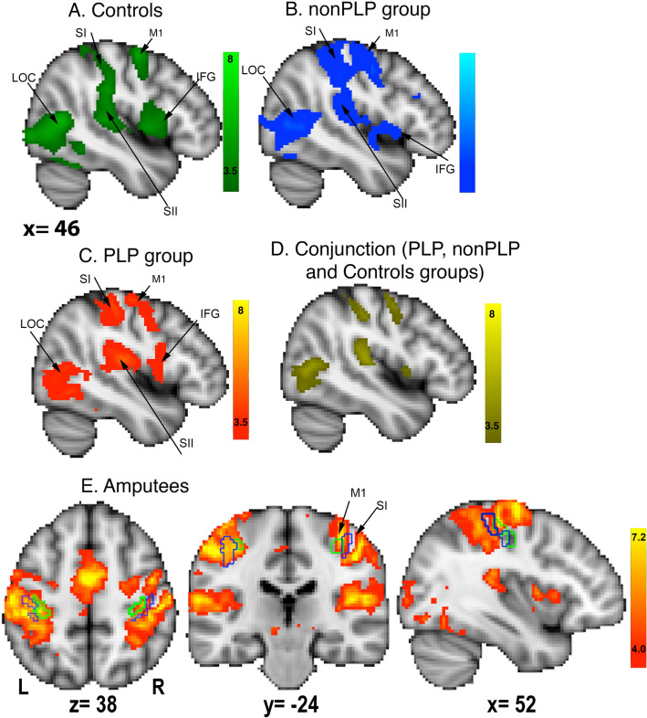
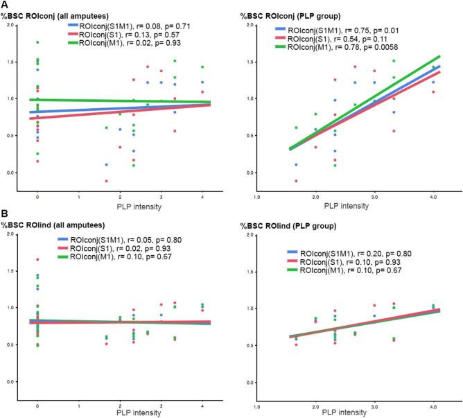
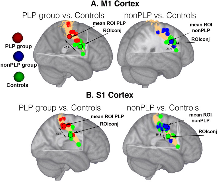
Phantom limb pain (PLP) has been associated with reorganization in primary somatosensory cortex (S1) and preserved S1 function. Here we examined if methodological differences in the assessment of cortical representations might explain these findings. We used functional magnetic resonance imaging during a virtual reality movement task, analogous to the classical mirror box task, in twenty amputees with and without PLP and twenty matched healthy controls. We assessed the relationship between task-related activation maxima and PLP intensity in S1 and motor cortex (M1) in individually-defined or group-conjoint regions of interest (ROI) (overlap of task-related activation between the groups). We also measured cortical distances between both locations and correlated them with PLP intensity. Amputees compared to controls showed significantly increased activation in M1, S1 and S1M1 unrelated to PLP. Neural activity in M1 was positively related to PLP intensity in amputees with PLP when a group-conjoint ROI was chosen. The location of activation maxima differed between groups in S1 and M1. Cortical distance measures were unrelated to PLP. These findings suggest that sensory and motor maps differentially relate to PLP and that methodological differences might explain discrepant findings in the literature.


Conflict of interest statement

The authors declare no competing interests.

Figures

Figure 1
Figure 1
Mean task-related activity during the virtual phantom task in the hemisphere contralateral to amputation (matched in healthy controls) for a the control group (green-lightgreen), b the nonPLP group (blue-lightblue) and c the PLP group (red-yellow). d Conjunction of task-related neural activity between the controls, PLP, and nonPLP groups (yellow). e Mean neural activity during the virtual phantom task in the amputee group. The green contours indicate the borders of the primary motor cortex (BA4p) and the blue contours indicate the borders for the primary somatosensory cortex (BA3b) in both hemispheres based on Juelich histological atlas. Activations are mapped on a MNI152 template provided by FSL, with FWE p < 0.05, TFCE, z > 2.3 threshold. Abbreviations: SI: primary somatosensory cortex, M1: primary motor cortex, LOC: lateral occipital cortex, IFG: inferior frontal gyrus, SII: secondary somatosensory cortex.
Figure 2
Figure 2
Correlation analyses between PLP intensity and %BSC in S1 (red), M1 (green) and S1M1 (blue) areas using a ROIconj and b ROIind in the entire sample of amputees (left) and in the PLP group only (right).
Figure 3
Figure 3
a Individual ROIs based on peak coordinates for the right hand ROI for the PLP (red), the nonPLP group (blue) and controls (green) in a the M1 cortex and b S1 cortex. The ROI for the controls overlapped entirely with ROIconj. The ROIs with a black contour indicate the mean of the individual ROIs. The surface coloured in copper indicates the mask of M1 (a) and S1 (b) defined by the Juelich atlas. Double-headed arrows indicate cortical distances in mm. ROIs are mapped on a MNI152 template provided by FSL.

References

    1. Elbert T, Pantev C, Wienbruch C, Rockstroh B, Taub E. Increased cortical representation of the fingers of the left hand in string players. Science. 1995;270:305–307. doi: 10.1126/science.270.5234.305. - DOI - PubMed
    1. Molina-Luna K, Hertler B, Buitrago MM, Luft AR. Motor learning transiently changes cortical somatotopy. Neuroimage. 2008;40:1748–1754. doi: 10.1016/j.neuroimage.2007.11.018. - DOI - PubMed
    1. Blume KR, et al. Cortical reorganization after macroreplantation at the upper extremity: a magnetoencephalographic study. Brain. 2014;137:757–769. doi: 10.1093/brain/awt366. - DOI - PubMed
    1. Langer N, Hanggi J, Muller NA, Simmen HP, Jancke L. Effects of limb immobilization on brain plasticity. Neurology. 2012;78:182–188. doi: 10.1212/WNL.0b013e31823fcd9c. - DOI - PubMed
    1. Pons TP, et al. Massive cortical reorganization after sensory deafferentation in adult macaques. Science. 1991;252:1857–1860. doi: 10.1126/science.1843843. - DOI - PubMed

Publication types