Review
. 2020 Nov;22(11):1743-1757.
doi: 10.1038/s41436-020-0877-3. Epub 2020 Jul 14.

A six-attribute classification of genetic mosaicism

Affiliations
Review

A six-attribute classification of genetic mosaicism

Víctor Martínez-Glez et al. Genet Med. 2020 Nov.

Abstract

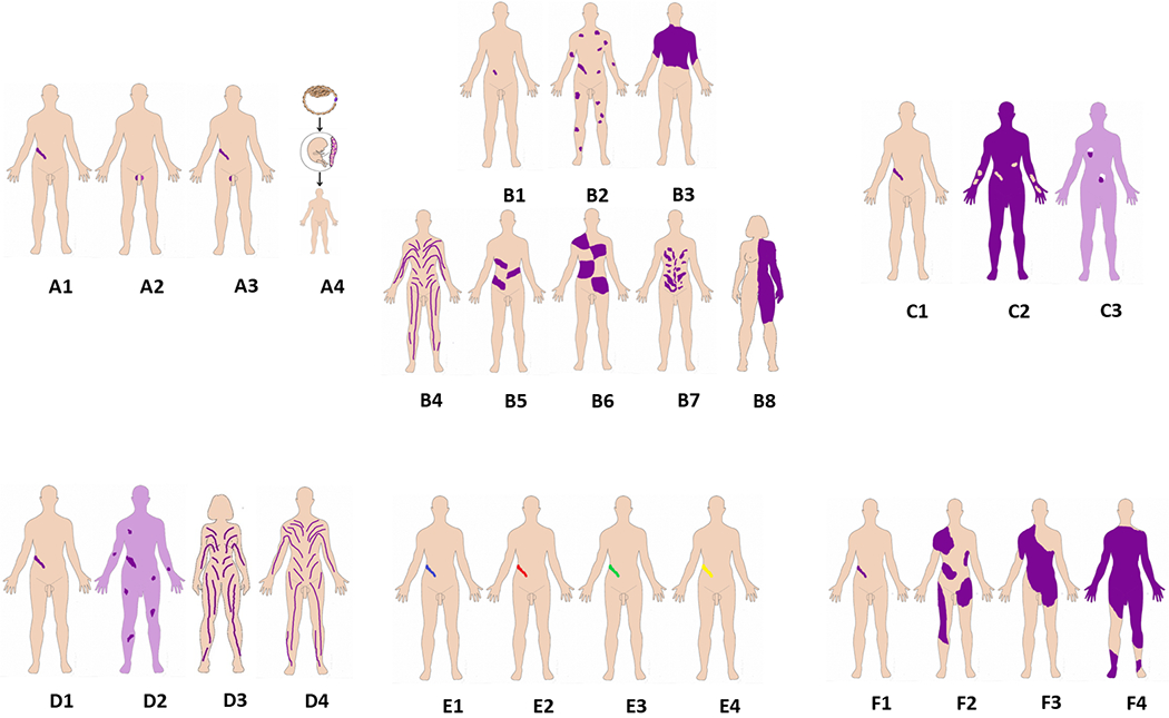
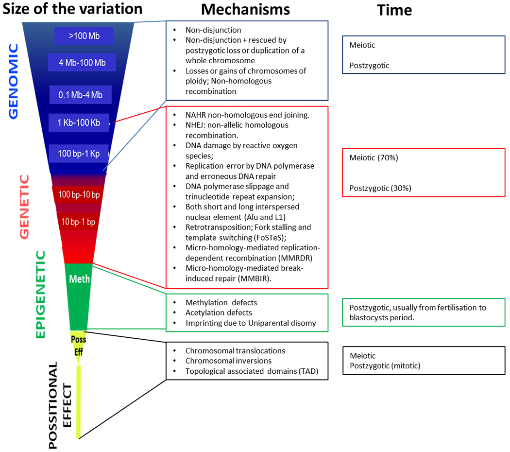
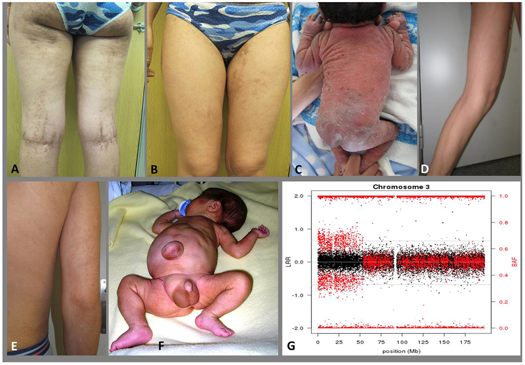
Mosaicism denotes an individual who has at least two populations of cells with distinct genotypes that are derived from a single fertilized egg. Genetic variation among the cell lines can involve whole chromosomes, structural or copy-number variants, small or single-nucleotide variants, or epigenetic variants. The mutational events that underlie mosaic variants occur during mitotic cell divisions after fertilization and zygote formation. The initiating mutational event can occur in any types of cell at any time in development, leading to enormous variation in the distribution and phenotypic effect of mosaicism. A number of classification proposals have been put forward to classify genetic mosaicism into categories based on the location, pattern, and mechanisms of the disease. We here propose a new classification of genetic mosaicism that considers the affected tissue, the pattern and distribution of the mosaicism, the pathogenicity of the variant, the direction of the change (benign to pathogenic vs. pathogenic to benign), and the postzygotic mutational mechanism. The accurate and comprehensive categorization and subtyping of mosaicisms is important and has potential clinical utility to define the natural history of these disorders, tailor follow-up frequency and interventions, estimate recurrence risks, and guide therapeutic decisions.

Keywords: mosaicism; mutational event; new classification; postzygotic.


Figures

Figure 1.
Figure 1.
Summary of the six-attribute classification of Genetic Mosaicism. The six attributes are illustrated in one panel each, A-F. Panel A, Affected tissues. A1: somatic mosaicism. A2: Germinal (gonadal) mosaicism. A3: Gonadosomatic mosaicism. A4: Confined placental mosaicism. Panel B, Body patterns. B1-B3 are non-segmented mosaicism. B1: single point mosaicism; B2: disseminated mosaicism; B3: patchy mosaicism without midline separation; B4-B8 are segmental mosaicism. B4. Blaschko lines in narrow bands; B5. Blaschko lines in broad bands; B6. Checkerboard; B7. Phylloid; B8. Lateralization/half body. Panel C, Change in Direction. C1: From benign (wild type) to pathogenic; C2: From pathogenic to benign (wild type): revertant mosaicism. C3: Didymosis. Panel D, Developmental mechanism. D1: Type 1 segmental, heterozygous state; D2: Type 2 segmental reflecting loss of the corresponding wild-type allele. D3: functional X-chromosome mosaicism with or without lethality for males; D4: Disorders due to autosomal lethal mutations that manifest only as mosaics. Panel E, Etiology. E1: genomic changes; E2: genetic changes; E3: epigenetic changes; E4: positional effects. Panel F, Fraction of the affected tissue. F1: Mild involvement; F2: Moderate involvement; F3: Severe involvement F4: Very severe (extreme) involvement (See also text and Suppl. Figures 1–6).
Figure 2.
Figure 2.
Scaled classes of variants (depending on the size of the variant, mechanism, and timing of the event). There are numerous endogenous molecular mechanisms that generate postzygotic somatic variants causing mosaicism during the lifetime of an individual, whereas others (such as Alu and L1 retrotransposition) are likely to have specific temporal patterns1, 37. The main genomic and epigenetic mechanisms are: a) DNA damage by reactive oxygen species; b) replication error by DNA polymerase and erroneous DNA repair; c) DNA polymerase slippage and trinucleotide repeat expansion; d) both short and long interspersed nuclear element (Alu and L1) retrotransposition; e) fork stalling and template switching (FoSTeS); f) non-homologous end joining (NHEJ); g) non-allelic homologous recombination (NAHR); h) micro-homology-mediated replication-dependent recombination (MMRDR); i) micro-homology-mediated break-induced repair (MMBIR); and j) losses or gains of chromosomes of ploidy, as reviewed in detail in 1 (See also Table S1). Exogenous factors such as tobacco and alcohol usage, and UV exposure may also be involved in postzygotic somatic mutational events37. We have divided these into four categories but recognize that the spectrum of variation is continuous, from a single nucleotide change to a diploid genome.
Figure 3:
Figure 3:
A and B: Epidermolytic epidermal nevus following Blaschko’s lines in a young woman and non-segmental epidermolytic ichthyosis of Brocq in her son (C). The mosaicism in the mother can be designated as: A3B4C1D1E2bF2: presumed to be gonadosomatic (A3), with a Blaschko pattern in narrow bands (B4), with a benign to pathogenic mutational direction of the variant (C1), type 1 postzygotic mechanism (heterozygous state) (D1), due to a genetic etiology (SNV in a gene (KRT1) (E2b) and between 10-30% of the tissue (skin) involved (F2) (see Suppl. material). D and E: Cutaneous mosaicism following the Blaschko lines in a young man. The pigmented lesions showed a trisomy 20 mosaicism (46,XY[24]/47,XY+20[14]). The mosaicism in this young man can be designated as: A1B4C1D1E1F1: a somatic mosaicism (A1), with a narrow lines of Blaschko pattern (B4), with a benign to pathogenic mutational direction of the variant (C1), type 1 postzygotic mechanism (heterozygous state) (D1), due to a genomic etiology (mosaic of chromosomes) (E1) and with mild involvement (<10%) of the tissue (skin) involved (F1) (see Suppl. material). F: A patient with Beckwith-Wiedemann syndrome and mosaic paternal uniparental disomy of the short arm of chromosome 11. Lateralized asymmetric overgrowth of the right side of the body, mainly in the leg. The mosaicism in this child can be designated as: A1B8C1D1E1fF3: a somatic mosaicism (A1), with a lateralization pattern (B8), with a healthy to pathogenic mutational direction of the variant (C1), a postzygotic mechanism (heterozygous state) (D1), due to an genomic etiology (mosaic of UPD of about 45 Mb) (E1f) and with moderate involvement (about 13%) of the tissue (the entire left leg) involved (F3) (see Suppl. material). G: SNP array plot detecting mosaic UPD 3p in peripheral blood DNA of a patient with Fanconi anemia. The chromosome 3 plot shows normal dosage along the entire chromosome (black dots with total intensity values – Log R Ratio-) and abnormal allelic dosage for the informative SNPs located in the distal 53 Mb of the p arm (red dots with relative allelic values –B Allelic Frequency-). Mosaicism in this child with an autosomal recessive disorder and a rescue mosaic UPD can be designated as: A1B0C2D1E1fF3: somatic mosaicism (A1), no pattern (B0), pathogenic to healthier (less pathogenic) mutational direction of the variant (C2), a postzygotic reversal mechanism (compound heterozygous to homozygous state) (D2), genomic etiology (UPD of 53 Mb) (E1f), involvement of about 33% (blood and buccal cells) (F3). (see Suppl. material).

References

    1. De S Somatic Mosaicism in Healthy Human Tissues. Trends in genetics : TIG. 2011;27(6):217–223. - PubMed
    1. Vera-Rodriguez M, Rubio C. Assessing the True Incidence of Mosaicism in Preimplantation Embryos. Fertility and sterility. 2017;107(5):1107–1112. - PubMed
    1. Happle R Mosaicism in Human Skin. 1 ed: Springer-Verlag Berlin Heidelberg; 2014.
    1. Bamford S, Dawson E, Forbes S, et al. The Cosmic (Catalogue of Somatic Mutations in Cancer) Database and Website. Br J Cancer. 2004;91(2):355–358. - PMC - PubMed
    1. Leija-Salazar M, Piette C, Proukakis C. Review: Somatic Mutations in Neurodegeneration. Neuropathology and applied neurobiology. 2018;44(3):267–285. - PubMed

LinkOut - more resources