Review
. 2021 Apr;48(2):187-198.
doi: 10.1007/s10396-020-01030-w. Epub 2020 Jul 13.

Role of endoscopic ultrasound for gallbladder disease

Affiliations
Review

Role of endoscopic ultrasound for gallbladder disease

Kazunari Tanaka et al. J Med Ultrason (2001). 2021 Apr.

Abstract

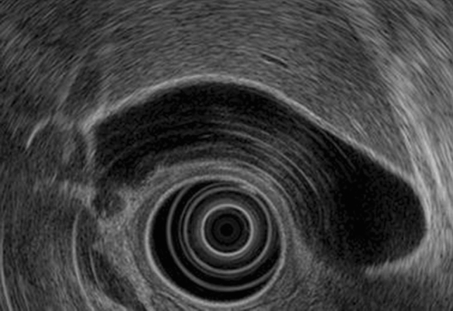
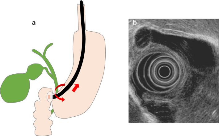
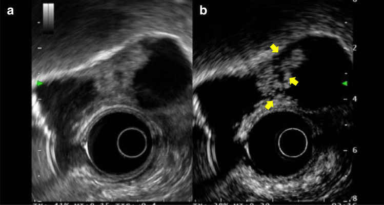
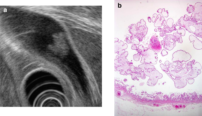
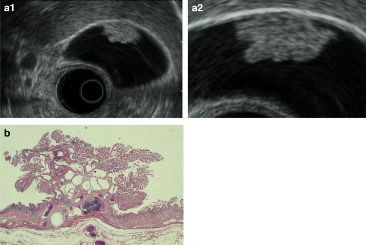
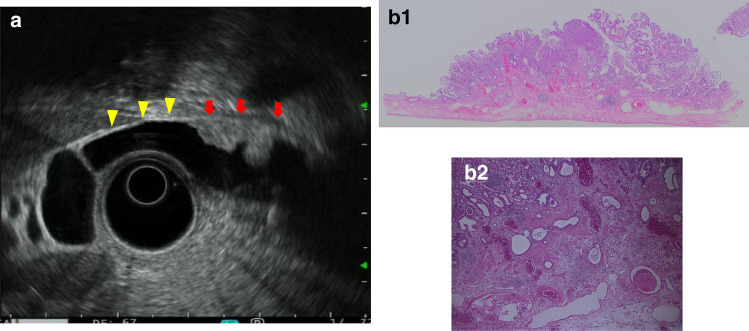
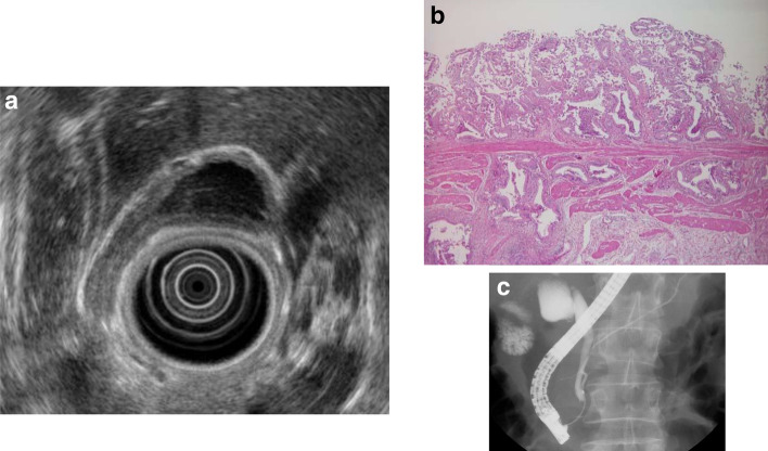
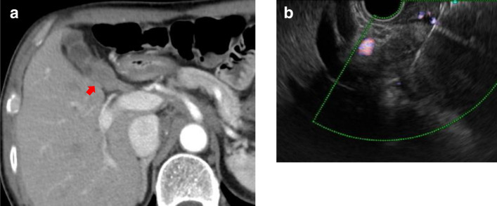
Endoscopic ultrasonography (EUS) has excellent spatial resolution and allows more detailed examination than abdominal ultrasonography (US) in terms of qualitative diagnosis of tumors and evaluation of tumor invasion depth. To understand the role of EUS in gallbladder disease, we need to understand the normal gallbladder wall structure and how to visualize it on EUS. In addition, gallbladder lesions can be classified into two broad categories: protuberant and wall-thickening lesions. Here, the features on EUS were outlined. We also outlined the current status of EUS-FNA for gallbladder lesions as there have been scattered reports of EUS-FNA in recent years.

Keywords: EUS; EUS-FNA; Gallbladder carcinoma; Gallbladder polyp.


Conflict of interest statement

The authors declare that there are no conflicts of interest.

Figures

Fig. 1
Fig. 1
Normal gallbladder. The gallbladder wall is divided into an inner low echoic layer and an outer high echoic layer
Fig. 2
Fig. 2
Radial scan type (short-scope position). a Short-scope position. b EUS image of the gallbladder in the short-scope position
Fig. 3
Fig. 3
Radial scan type (long-scope position). a Long-scope position in the duodenum bulb. b Clockwise rotation causes the scope to advance in the direction of the descending duodenum
Fig. 4
Fig. 4
Convex array (transgastric scanning). a EUS image of the cystic duct junction. b EUS image of the gallbladder neck and body. Gn: neck of gallbladder, Gb: body of gallbladder
Fig. 5
Fig. 5
Convex arrayed (duodenal bulb scanning). a EUS image of the cystic duct and gallbladder neck and body. b Fundus of gallbladder. Gf: fundus of gallbladder
Fig. 6
Fig. 6
Contrast EUS for gallbladder carcinoma. a Conventional EUS demonstrates a hypoechoic mass in the gallbladder. b Contrast-enhanced harmonic EUS indicates that the area has perfusion defects (arrow)
Fig. 7
Fig. 7
Cholesterol polyp. a This polyp has a granular surface and morular morphology. The internal echo is rough or granular. b Polypoid lesion with non-neoplastic epithelium and abundant stroma
Fig. 8
Fig. 8
Cholesterol polyp resembling early gallbladder carcinoma. a EUS image of a solid internal echogenicity polyp without echogenic punctiform foci. b (1, 2) Photomicrograph demonstrating an aggregation of foamy cells under the epithelium
Fig. 9
Fig. 9
Gallbladder hyperplastic polyp. a EUS image of a pedunculated, lobulated, solid internal echogenicity polyp. b (1, 2) The polyp consists of duct glands similar to the pyloric gland
Fig. 10
Fig. 10
Gallbladder inflammatory polyp. a EUS image showing a pedunculated, smooth surface polyp with an anechoic area. b (1, 2) The stroma consists of edematous and coarse fibrous connective tissue. The surface iconsists of simple columnar epithelium
Fig. 11
Fig. 11
Gallbladder fibrous polyp. a EUS image showing a pedunculated, smooth surface, uniformal echogenicity hypoechoic polyp. b (1, 2) The stroma consists of edematous and coarse fibrous connective tissue. The surface consists of simple columnar epithelium
Fig. 12
Fig. 12
Gallbladder adenoma. a (1, 2) EUS image showing a relatively smooth surface, solid internal echogenicity polyp with multiple microcystic spaces. b Photomicrograph imaging of the gallbladder adenoma
Fig. 13
Fig. 13
Early gallbladder carcinoma. a (1, 2) EUS image: A homogenously hypoechoic protruding lesion with a granular surface is seen. The outer layer of the gallbladder is well preserved (arrow, retained hyperechoic outer layer; arrowhead, normal hyperechoic outer layer). b Photomicrograph: A pedunculated polypoid lesion was diagnosed as well-differentiated adenocarcinoma. It was invading into but not through the muscularis layer
Fig. 14
Fig. 14
Advanced gallbladder carcinoma. a EUS imaging of a sessile elevated lesion with thinning of the hyperechoic outer layer (arrow, thinning of the hyperechoic outer layer; arrowhead, normal hyperechoic outer layer). b (1, 2) Photomicrograph: Gallbladder carcinoma with SS depth invasion
Fig. 15
Fig. 15
Gallbladder adenomyomatosis. a Fundal type. b Diffuse type. c Segmental type
Fig. 16
Fig. 16
Hyperplasia of the gallbladder mucous membrane accompanying anomalous pancreaticobiliary junction. a EUS image of the thickened inner hypoechoic layer of the gallbladder. b Hyperplastic changes in the gallbladder mucous membrane. c ERCP image of anomalous pancreaticobiliary junction
Fig. 17
Fig. 17
Gallbladder carcinoma (wall-thickening type). a (1, 2) EUS image of irregular gallbladder wall thickening from the gallbladder body to the fundus (arrow). b (1, 2) Photomicrograph: Gallbladder carcinoma with SS depth invasion
Fig. 18
Fig. 18
EUS-FNA for a gallbladder lesions. a CT scan shows a gallbladder lesion in the gallbladder neck (arrow). b EUS-guided FNA for a gallbladder mass lesion

References

    1. Fujita N, Noda Y, Kobayashi G, et al. Analysis of the layer structure of the gallbladder wall delineated by endoscopic ultrasound using the pinning method. Dig Endosc. 1995;7:353–356. doi: 10.1111/j.1443-1661.1995.tb00387.x. - DOI
    1. Kaneko M, Katanuma A, Maguchi H. Prospective, randomized, comparative study of delineation capability of radial scanning and curved linear array endoscopic ultrasound for the pancreaticobiliary region. Endosc Int Open. 2014;2:E160–E170. doi: 10.1055/s-0034-1377384. - DOI - PMC - PubMed
    1. Choi JH, Seo DW, Choi JH, et al. Utility of contrast-enhanced harmonic EUS in the diagnosis of malignant gallbladder polyps. Gastrointest Endosc. 2013;78:484–493. doi: 10.1016/j.gie.2013.03.1328. - DOI - PubMed
    1. Imazu H, Mori N, Tajiri H, et al. Contrast-enhanced harmonic endoscopic ultrasonography in the differential diagnosis of gallbladder wall thickening. Dig Dis Sci. 2014;59:1909–1916. doi: 10.1007/s10620-014-3115-5. - DOI - PubMed
    1. Karaosmmanoglu AD, Blake M. Hamartomatous polyp of the gallbladder with an associated choledochal cyst. J Ultrasound Med. 2010;29:1663–1666. doi: 10.7863/jum.2010.29.11.1663. - DOI - PubMed

MeSH terms