Review
. 2020 Aug;12(4):1007-1017.
doi: 10.1007/s12551-020-00729-x. Epub 2020 Jul 13.

The architecture and function of cardiac dyads

Affiliations
Review

The architecture and function of cardiac dyads

Fujian Lu et al. Biophys Rev. 2020 Aug.

Abstract

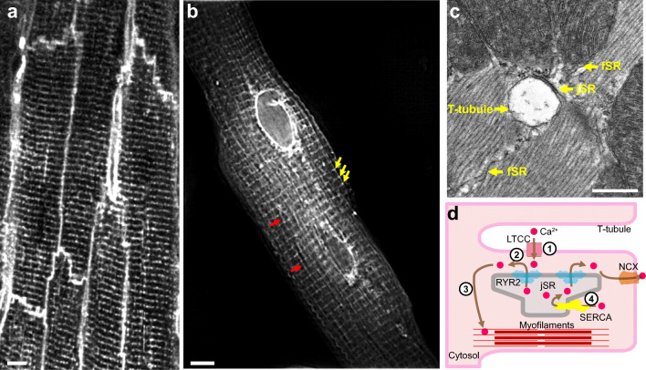
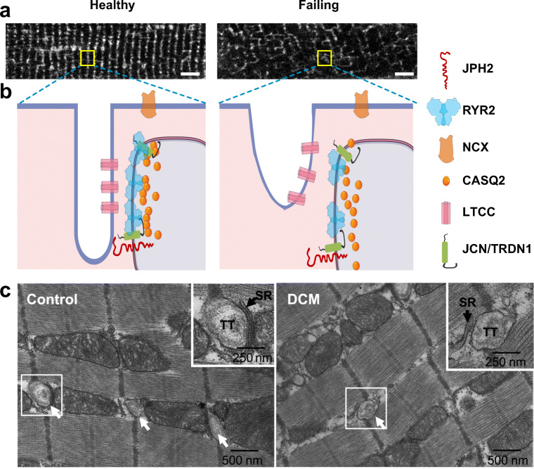
Cardiac excitation-contraction (EC) coupling, which links plasma membrane depolarization to activation of cardiomyocyte contraction, occurs at dyads, the nanoscopic microdomains formed by apposition of transverse (T)-tubules and junctional sarcoplasmic reticulum (jSR). In a dyadic junction, EC coupling occurs through Ca2+-induced Ca2+ release. Membrane depolarization opens voltage-gated L-type Ca2+ channels (LTCCs) in the T-tubule. The resulting influx of extracellular Ca2+ into the dyadic cleft opens Ca2+ release channels known as ryanodine receptors (RYRs) in the jSR, leading to the rapid increase in cytosolic Ca2+ that triggers sarcomere contraction. The efficacy of LTCC-RYR communication greatly affects a myriad of downstream intracellular signaling events, and it is controlled by many factors, including T-tubule and jSR structure, spatial distribution of ion channels, and regulatory proteins that closely regulate the activities of channels within dyads. Alterations in dyad architecture and/or channel activity are seen in many types of heart disease. This review will focus on the current knowledge regarding cardiac dyad structure and function, their alterations in heart failure, and new approaches to study the composition and function of dyads.

Keywords: Cardiac dyads; EC coupling; T-tubule; jSR.


Conflict of interest statement

The authors declare that they have no conflicts of interest.

Figures

Fig. 1
Fig. 1
Anatomy of cardiac dyads. (A) T-tubule network of mature adult cardiomyocytes. The mouse heart was perfused with MM-4-64, which stains the sarcolemma, and optically sectioned and imaged using a confocal microscope. Bar = 5 μm. (B) Total sarcoplasmic reticulum (SR) structure of an adult cardiomyocyte. The dissociated adult mouse cardiomyocyte was stained using Fluo5N and visualized by structured illumination microscopy. Note the discrete junctional SR (jSR; yellow arrows) and the diffusible free SR (fSR; red arrows) and the much brighter perinuclear envelopes. Bar = 5 μm. (C) Longitudinal section of mouse left ventricular myocyte in a thin section transmission electron micrograph. A T-tubule, shown in cross section, is separated from pancake-shaped jSR cisternae by a narrow (~12 nm) junctional gap. Electron dense “foot” structures corresponding to the cytoplasmic domain of RYR2 protrude into the gap. fSR is contiguous with jSR. Bar = 0.5 μm. (D) Cartoon showing the process of EC coupling. At dyadic junctions, L-type Ca2+ channels (LTCCs) in T-tubules are coupled with RYR2 Ca2+ release channels in jSR. (1) The action potential depolarizes T-tubule membranes, opening LTCCs. (2) Extracellular Ca2+ enters through LTCCs into dyadic clefts, resulting in Ca2+-induced Ca2+ release through RYR2. (3) The resulting increase in [Ca2+]i causes sarcomere contraction. (4) [Ca2+]i is returned to basal levels by SERCA2a, which actively pumps Ca2+ back into SR, and by extrusion through the Na+/Ca2+ exchanger, NCX
Fig. 2
Fig. 2
Altered dyad architecture in heart failure. (A) Confocal micrographs of the T-tubule system revealed by MM4-64 membrane dye in healthy and diseased murine cardiomyocytes. Note the T-tubular disorganization in the diseased cardiomyocyte. (B) Schematic illustration of normal (left) and diseased (right) dyadic junctions. The diseased dyad architecture features T-tubule loss and remodeling, ion channel dislocation, junctional contact area shrinkage, LTCC-RYR2 uncoupling, RYR2 cluster redistribution, and disordered fuzzy space (see text). Bar = 5 μm. JCN, junctin; TRDN1, triadin 1. (C) Transmission electron micrographs of human LV myocardium from healthy control (left) or patient with dilated cardiomyopathy (DCM, right).Arrows point to T-tubules (TT). Boxed areas are enlarged in upper right corner. In control, jSR is separated from T-tubule by a narrow junctional gap. In DCM, the junctional gap is wider, and the contact area is reduced. Panel C was reproduced from Zhang et al. (2013) with permission from the publisher
Fig. 3
Fig. 3
New approaches to dissecting the structure and functional regulation of dyads. (A–B) Proximity proteomics applied to probe the dyadic proteome. Purple-shaded regions indicate labeling sphere determined by biotin free radical half-life. (A) APEX2-based proximity proteomics. Liu et al. (2020) fused APEX2 to components of the LTCC. Quantitative proteomics showed that β-adrenergic signaling regulated LTCC activity by controlling interaction of the LTCC β2B subunit with the novel dyad protein RAD. (B) Feng et al. (2020) fused endogenous JPH2 to BioID2 to detect nearby dyadic proteins. (C–E) Cas9 and AAV (CASAAV) strategy to generate somatic mutations in cardiomyocytes. AAV expressing gRNAs that target the gene of interest and cardiomyocyte-restricted Cre is administered to mice with genome-encoded, Cre-activated Cas9-P2A-GFP. Cre activates Cas9 and GFP, and Cas9 plus gRNAs inactivate the gene of interest. (D) Inactivation of JPH2 in Cas9-P2A-GFP expressing cardiomyocytes. Mice were treated with CASAAV vector targeting Jph2. At P21, dissociated cardiomyocytes were imaged to detect GFP and JPH2. Note the loss of JPH2 immunoreactivity in GFP+ cardiomyocyte (arrow). (E) Mosaic gene inactivation by CASAAV permits precise interpretation of results. CASAAV vector targeting JPH2 was given at lower or high doses. High dose, which caused heart failure, resulted in distorted T-tubule morphology in GFP+ and GFP− cells. Low dose, which did not cause heart failure, did not visibly disrupt T-tubules in GFP+ or GFP− cells. D–E were modified from Guo et al. (2017) with permission

References

    1. Acsai K, Antoons G, Livshitz L, et al. Microdomain [Ca2+] near ryanodine receptors as reported by L-type Ca2+ and Na+/Ca2+ exchange currents. J Physiol. 2011;589:2569–2583. - PMC - PubMed
    1. Baddeley D, Jayasinghe ID, Lam L, et al. Optical single-channel resolution imaging of the ryanodine receptor distribution in rat cardiac myocytes. Proc Natl Acad Sci. 2009;106:22275–22280. - PMC - PubMed
    1. Bers D (2001) Excitation-contraction coupling and cardiac contractile force. Springer Science & Business Media
    1. Bers DM. Cardiac excitation–contraction coupling. Nature. 2002;415:198–205. - PubMed
    1. Bongianino R, Denegri M, Mazzanti A, et al. Allele-specific silencing of mutant mRNA rescues ultrastructural and arrhythmic phenotype in mice carriers of the R4496C mutation in the ryanodine receptor gene (RYR2) Circ Res. 2017;121:525–536. - PubMed

LinkOut - more resources