

Does the human placenta express the canonical cell entry mediators for SARS-CoV-2?
- PMID: 32662421
- PMCID: PMC7367681
- DOI: 10.7554/eLife.58716
Does the human placenta express the canonical cell entry mediators for SARS-CoV-2?
Abstract
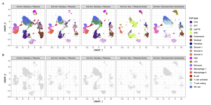
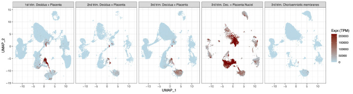
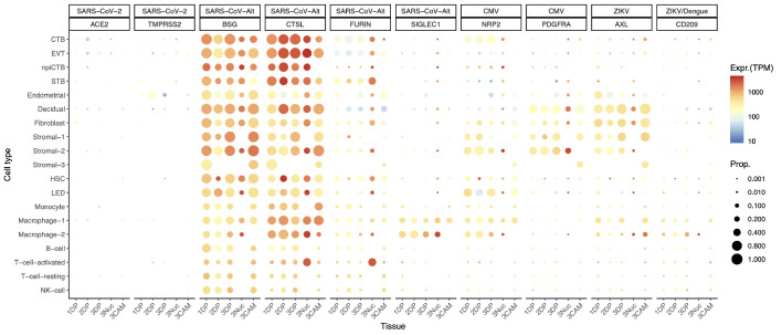
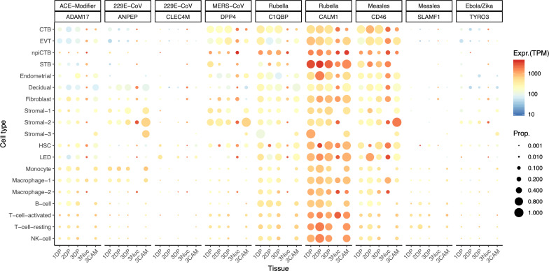
The pandemic of coronavirus disease 2019 (COVID-19) caused by the severe acute respiratory syndrome coronavirus 2 (SARS-CoV-2) has affected more than 10 million people, including pregnant women. To date, no consistent evidence for the vertical transmission of SARS-CoV-2 exists. The novel coronavirus canonically utilizes the angiotensin-converting enzyme 2 (ACE2) receptor and the serine protease TMPRSS2 for cell entry. Herein, building upon our previous single-cell study (Pique-Regi et al., 2019), another study, and new single-cell/nuclei RNA-sequencing data, we investigated the expression of ACE2 and TMPRSS2 throughout pregnancy in the placenta as well as in third-trimester chorioamniotic membranes. We report that co-transcription of ACE2 and TMPRSS2 is negligible in the placenta, thus not a likely path of vertical transmission for SARS-CoV-2. By contrast, receptors for Zika virus and cytomegalovirus, which cause congenital infections, are highly expressed by placental cell types. These data show that the placenta minimally expresses the canonical cell-entry mediators for SARS-CoV-2.
Keywords: COVID-19; SARS-CoV-2; genetics; genomics; human; human biology; medicine; placenta; pregnancy vertical transmisssion; scRNA-seq; viral receptors.
Conflict of interest statement
RP, RR, AT, FL, YX, AA, YL, CH, NG No competing interests declared
Figures
References
-
- Aagaard KM, Lahon A, Suter MA, Arya RP, Seferovic MD, Vogt MB, Hu M, Stossi F, Mancini MA, Harris RA, Kahr M, Eppes C, Rac M, Belfort MA, Park CS, Lacorazza D, Rico-Hesse R. Primary human placental trophoblasts are permissive for zika virus (ZIKV) Replication. Scientific Reports. 2017;7:41389. doi: 10.1038/srep41389. - DOI - PMC - PubMed
-
- Adams Waldorf KM, Stencel-Baerenwald JE, Kapur RP, Studholme C, Boldenow E, Vornhagen J, Baldessari A, Dighe MK, Thiel J, Merillat S, Armistead B, Tisoncik-Go J, Green RR, Davis MA, Dewey EC, Fairgrieve MR, Gatenby JC, Richards T, Garden GA, Diamond MS, Juul SE, Grant RF, Kuller L, Shaw DW, Ogle J, Gough GM, Lee W, English C, Hevner RF, Dobyns WB, Gale M, Rajagopal L. Fetal brain lesions after subcutaneous inoculation of zika virus in a pregnant nonhuman primate. Nature Medicine. 2016;22:1256–1259. doi: 10.1038/nm.4193. - DOI - PMC - PubMed
-
- Adams Waldorf KM, Nelson BR, Stencel-Baerenwald JE, Studholme C, Kapur RP, Armistead B, Walker CL, Merillat S, Vornhagen J, Tisoncik-Go J, Baldessari A, Coleman M, Dighe MK, Shaw DWW, Roby JA, Santana-Ufret V, Boldenow E, Li J, Gao X, Davis MA, Swanstrom JA, Jensen K, Widman DG, Baric RS, Medwid JT, Hanley KA, Ogle J, Gough GM, Lee W, English C, Durning WM, Thiel J, Gatenby C, Dewey EC, Fairgrieve MR, Hodge RD, Grant RF, Kuller L, Dobyns WB, Hevner RF, Gale M, Rajagopal L. Congenital zika virus infection as a silent pathology with loss of neurogenic output in the fetal brain. Nature Medicine. 2018;24:368–374. doi: 10.1038/nm.4485. - DOI - PMC - PubMed
-
- Al-Haddad BJS, Oler E, Armistead B, Elsayed NA, Weinberger DR, Bernier R, Burd I, Kapur R, Jacobsson B, Wang C, Mysorekar I, Rajagopal L, Adams Waldorf KM. The fetal origins of mental illness. American Journal of Obstetrics and Gynecology. 2019;221:549–562. doi: 10.1016/j.ajog.2019.06.013. - DOI - PMC - PubMed
Publication types
MeSH terms
Substances
Associated data
Grants and funding
LinkOut - more resources
Full Text Sources
Molecular Biology Databases
Miscellaneous
