

Ultrashort echo time quantitative susceptibility mapping (UTE-QSM) for detection of hemosiderin deposition in hemophilic arthropathy: A feasibility study
- PMID: 32662904
- PMCID: PMC7895482
- DOI: 10.1002/mrm.28388
Ultrashort echo time quantitative susceptibility mapping (UTE-QSM) for detection of hemosiderin deposition in hemophilic arthropathy: A feasibility study
Abstract
Purpose: The purpose of this study was to investigate the feasibility of ultrashort echo time quantitative susceptibility mapping (UTE-QSM) for assessment of hemosiderin deposition in the joints of hemophilic patients.
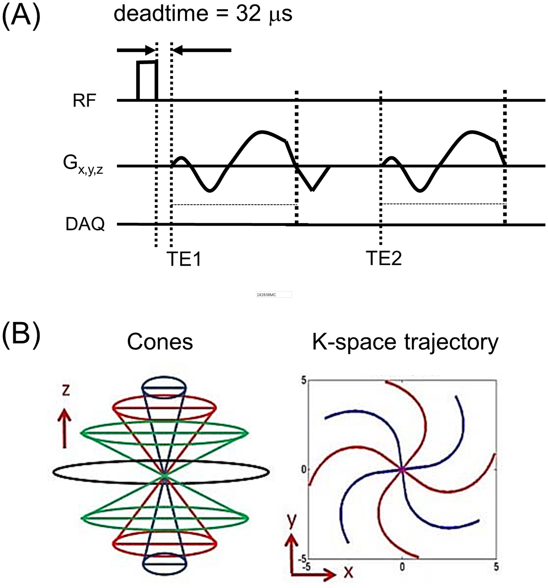
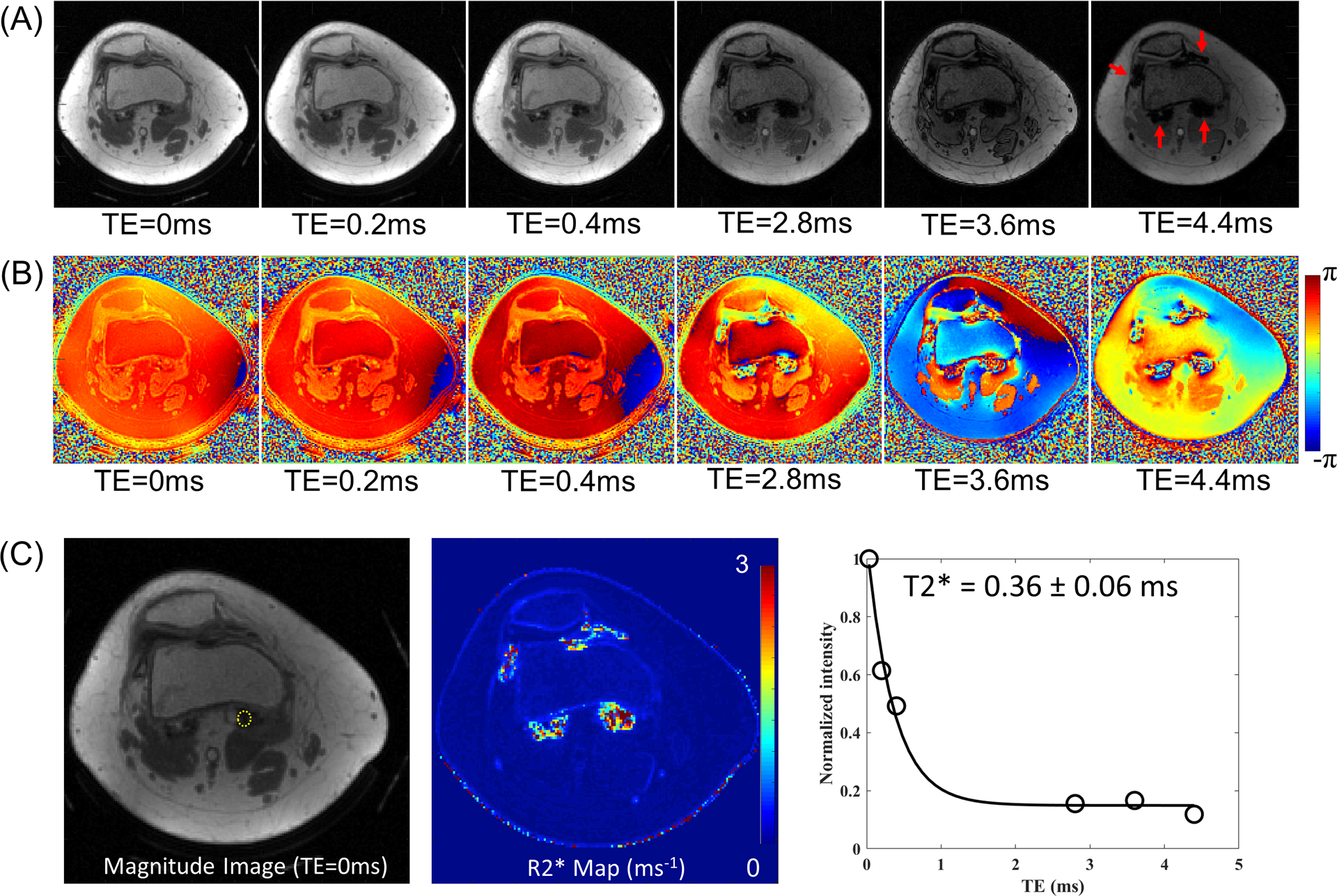
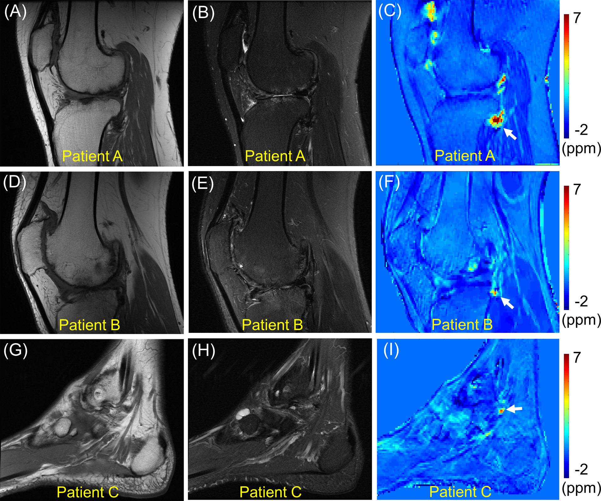
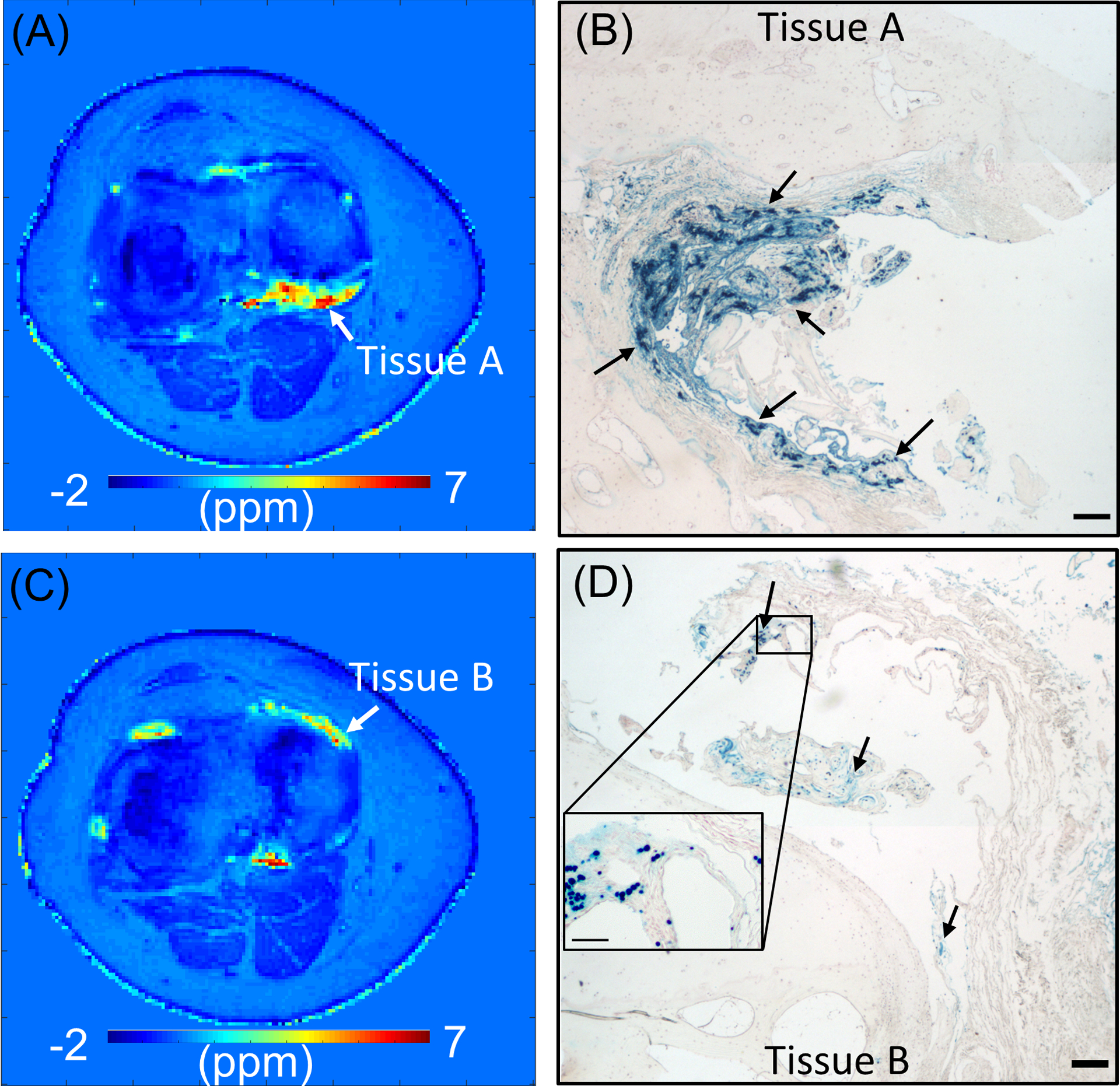
Methods: The UTE-QSM technique was based on three sets of dual-echo 3D UTE Cones data acquired with TEs of 0.032/2.8, 0.2/3.6, and 0.4/4.4 ms. The images were processed with iterative decomposition of water and fat with echo asymmetry and least-squares estimation to estimate the B0 field map in the presence of fat. Then, the projection onto dipole field (PDF) algorithm was applied to acquire a local field map generated by tissues, followed by application of the morphology-enabled dipole inversion (MEDI) algorithm to estimate a final susceptibility map. Three healthy volunteers and three hemophilic patients were recruited to evaluate the UTE-QSM technique's ability to assess hemosiderin in the knee or ankle joint at 3T. One patient subsequently underwent total knee arthroplasty after the MR scan. The synovial tissues harvested from the knee joint during surgery were processed for histological analysis to confirm iron deposition.
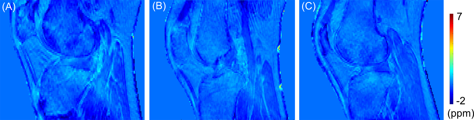
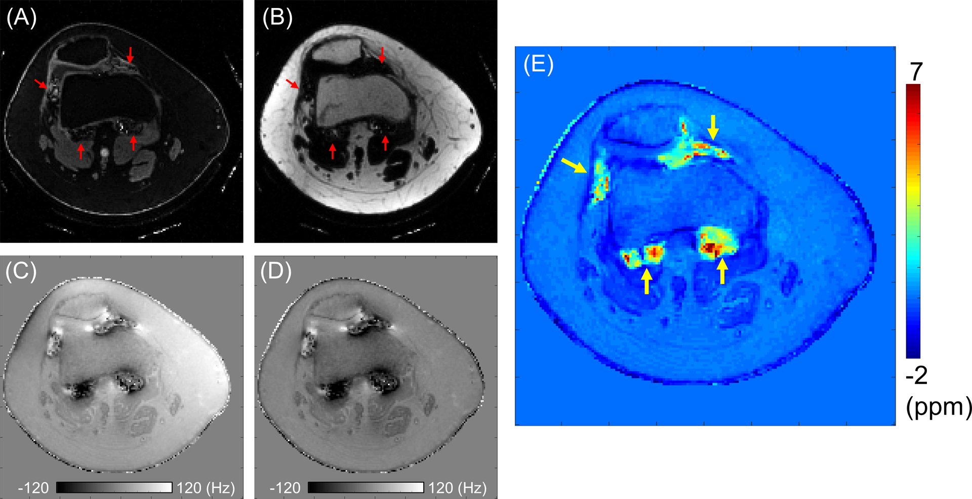
Results: UTE-QSM successfully yielded tissue susceptibility maps of joints in both volunteers and patients. Multiple regions with high susceptibility over 1 ppm were detected in the affected joints of hemophilic patients, while no localized regions with high susceptibility were detected in asymptomatic healthy volunteers. Histology confirmed the presence of iron in regions where high susceptibility was detected by UTE-QSM.
Conclusion: The UTE-QSM technique can detect hemosiderin deposition in the joint, and provides a potential sensitive biomarker for the diagnosis and prognosis of hemophilic arthropathy.
Keywords: QSM; UTE; ankle; arthropathy; hemophilia; knee; susceptibility.
© 2020 International Society for Magnetic Resonance in Medicine.
Figures
Similar articles
-
Hemosiderin quantification in hemophilic arthropathy using quantitative magnetic resonance imaging.Sci Rep. 2025 Apr 24;15(1):14353. doi: 10.1038/s41598-025-99085-7. Sci Rep. 2025. PMID: 40274909 Free PMC article.
-
Feasibility of ultrashort echo time quantitative susceptibility mapping with a 3D cones trajectory in the human brain.Front Neurosci. 2022 Nov 7;16:1033801. doi: 10.3389/fnins.2022.1033801. eCollection 2022. Front Neurosci. 2022. PMID: 36419458 Free PMC article.
-
Simultaneous quantitative susceptibility mapping (QSM) and R2* for high iron concentration quantification with 3D ultrashort echo time sequences: An echo dependence study.Magn Reson Med. 2018 Apr;79(4):2315-2322. doi: 10.1002/mrm.27062. Epub 2018 Jan 4. Magn Reson Med. 2018. PMID: 29314215 Free PMC article.
-
Early differentiation of neurodegenerative diseases using the novel QSM technique: what is the biomarker of each disorder?BMC Neurosci. 2022 Jul 28;23(1):48. doi: 10.1186/s12868-022-00725-9. BMC Neurosci. 2022. PMID: 35902793 Free PMC article. Review.
-
Quantitative Susceptibility Mapping: Basic Methods and Clinical Applications.Radiographics. 2022 Jul-Aug;42(4):1161-1176. doi: 10.1148/rg.210054. Epub 2022 May 6. Radiographics. 2022. PMID: 35522577 Review.
Cited by
-
QSM Throughout the Body.J Magn Reson Imaging. 2023 Jun;57(6):1621-1640. doi: 10.1002/jmri.28624. Epub 2023 Feb 7. J Magn Reson Imaging. 2023. PMID: 36748806 Free PMC article. Review.
-
Making the invisible visible-ultrashort echo time magnetic resonance imaging: Technical developments and applications.Appl Phys Rev. 2022 Dec;9(4):041303. doi: 10.1063/5.0086459. Appl Phys Rev. 2022. PMID: 36467869 Free PMC article. Review.
-
Ultrashort echo time Cones double echo steady state (UTE-Cones-DESS) for rapid morphological imaging of short T2 tissues.Magn Reson Med. 2021 Aug;86(2):881-892. doi: 10.1002/mrm.28769. Epub 2021 Mar 23. Magn Reson Med. 2021. PMID: 33755258 Free PMC article.
-
Deep Convolutional Neural Network for Dedicated Regions-of-Interest Based Multi-Parameter Quantitative Ultrashort Echo Time (UTE) Magnetic Resonance Imaging of the Knee Joint.J Imaging Inform Med. 2024 Oct;37(5):2126-2134. doi: 10.1007/s10278-024-01089-8. Epub 2024 Mar 28. J Imaging Inform Med. 2024. PMID: 38548992 Free PMC article.
-
Simultaneous Quantitative Susceptibility Mapping of Articular Cartilage and Cortical Bone of Human Knee Joint Using Ultrashort Echo Time Sequences.Front Endocrinol (Lausanne). 2022 Feb 22;13:844351. doi: 10.3389/fendo.2022.844351. eCollection 2022. Front Endocrinol (Lausanne). 2022. PMID: 35273576 Free PMC article.
References
-
- Kidder W, Nguyen S, Larios J, Bergstrom J, Ceponis A, von Drygalski A. Point-of-care musculoskeletal ultrasound is critical for the diagnosis of hemarthroses, inflammation and soft tissue abnormalities in adult patients with painful haemophilic arthropathy. Haemophilia 2015;21:530–537 doi: 10.1111/hae.12637. - DOI - PubMed
Publication types
MeSH terms
Substances
Grants and funding
LinkOut - more resources
Full Text Sources
Medical
