. 2020 Sep 2;11(17):2728-2740.
doi: 10.1021/acschemneuro.0c00363. Epub 2020 Aug 3.

Novel Curcumin-Diethyl Fumarate Hybrid as a Dualistic GSK-3β Inhibitor/Nrf2 Inducer for the Treatment of Parkinson's Disease

Affiliations

Novel Curcumin-Diethyl Fumarate Hybrid as a Dualistic GSK-3β Inhibitor/Nrf2 Inducer for the Treatment of Parkinson's Disease

Rita Maria Concetta Di Martino et al. ACS Chem Neurosci. .

Abstract

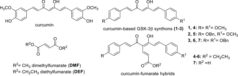
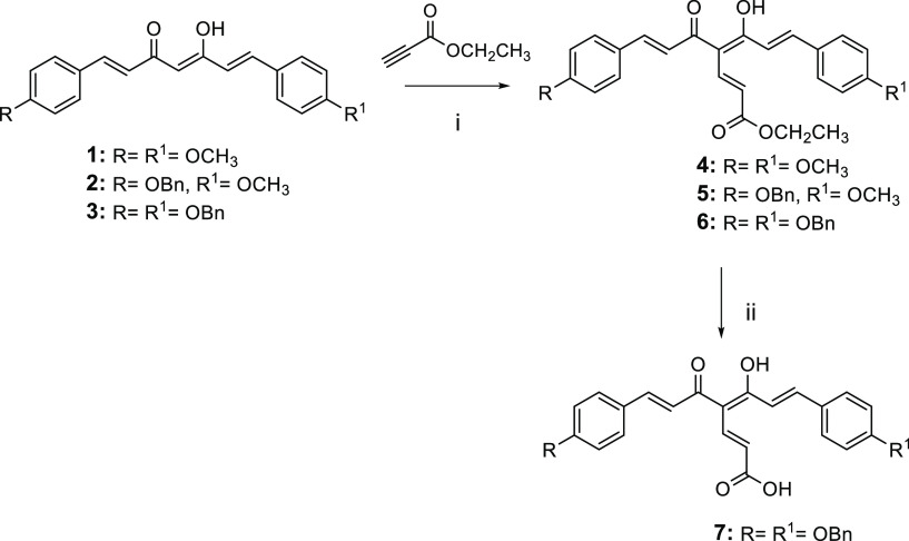
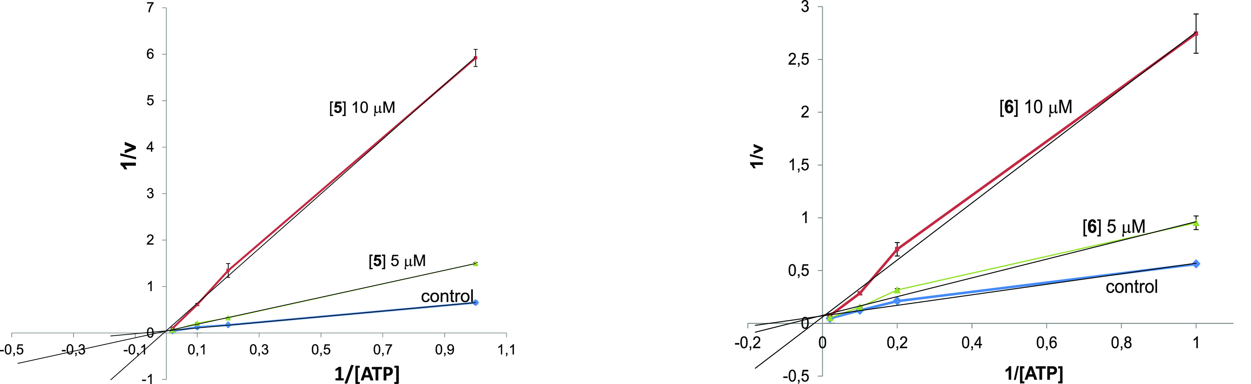
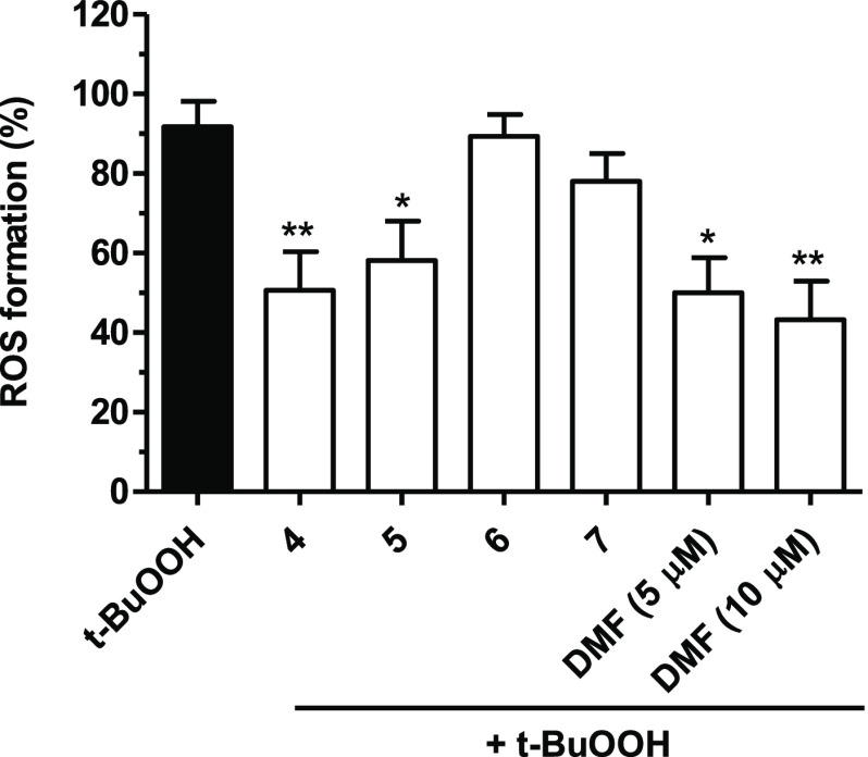
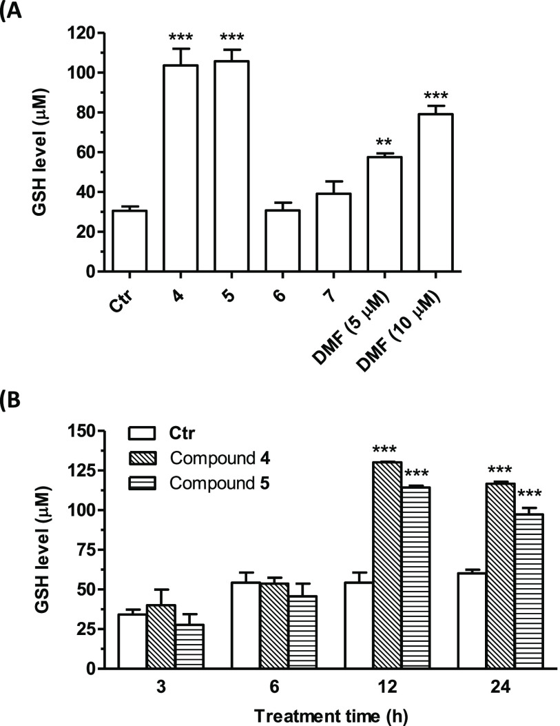
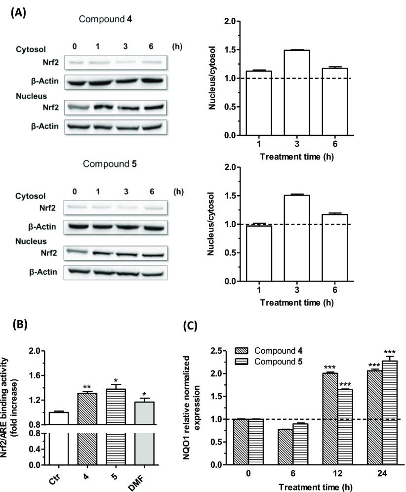
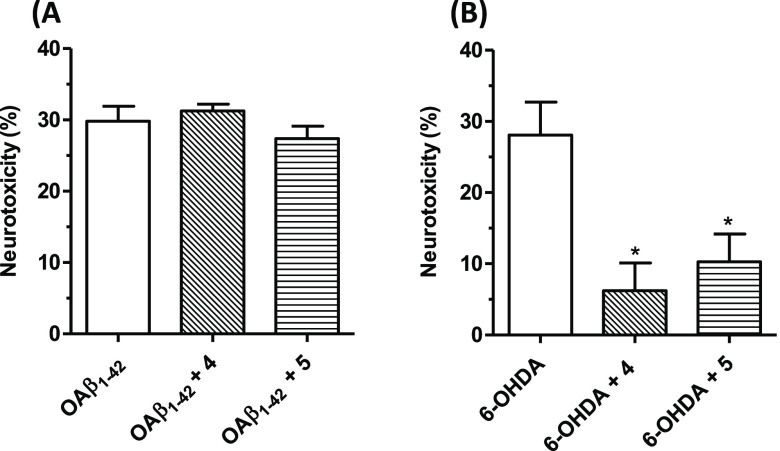
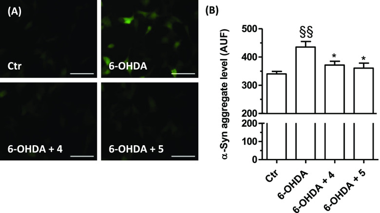
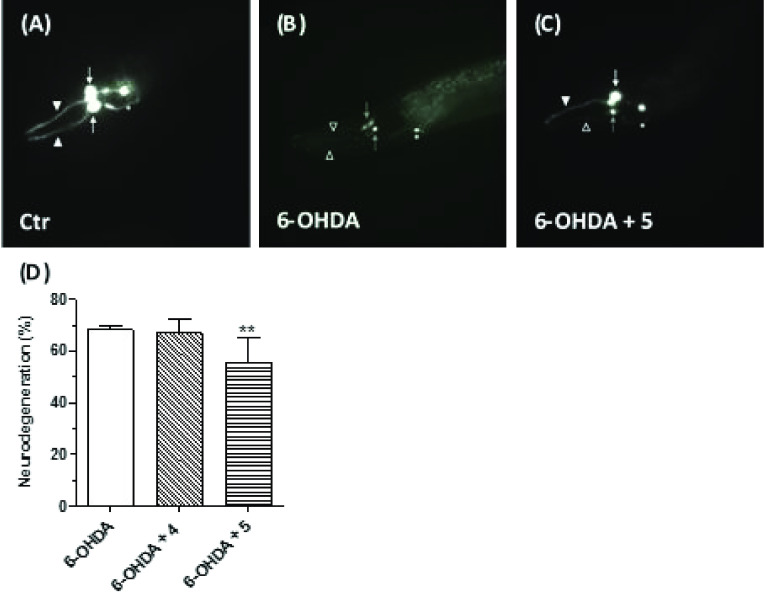
Common copathogenic factors, including oxidative stress and neuroinflammation, are found to play a vital role in the development of neurodegenerative disorders, including Alzheimer's disease (AD) and Parkinson's disease (PD). Nowadays, owing to the multifactorial character of the diseases, no effective therapies are available, thus underlying the need for new strategies. Overexpression of the enzyme GSK-3β and downregulation of the Nrf2/ARE pathway are responsible for a decrease in antioxidant defense effects. These pieces of evidence underline the usefulness of dual GSK-3β inhibitors/Nrf2 inducers. In this regard, to design a dual modulator, the structures of a curcumin-based analogue, as GSK-3β inhibitor, and a diethyl fumarate fragment, as Nrf2 inducer, were combined. Among the hybrids, 5 and 6 proved to effectively inhibit GSK-3β, while 4 and 5 showed a marked ability to activate Nrf2 together to increase the neuronal resistance to oxidative stress. These last pieces of evidence translated into specific neuroprotective effects of 4 and 5 against PD pathological events including neurotoxicity elicited by α-synuclein aggregates and 6-hydroxydopamine. Hybrid 5 also showed neuroprotective effects in a C. elegans model of PD where the activation of GSK-3β is intimately involved in Nrf2 regulation. In summary, 5 emerged as an interesting multitarget derivative, valuable to be exploited in a multitarget PD perspective.

Keywords: Curcumin analogues; Diethyl fumarate; Glycogen synthase kinase-3β; Neurodegeneration; Nuclear factor-erythroid related factor 2; Oxidative stress; Parkinson’s disease.


Conflict of interest statement

The authors declare no competing financial interest.

Figures

Figure 1
Figure 1
Chemical structures of curcumin and general structures of curcumin-based starting synthons (13), fumaric acid esters (DMF and DEF), and curcumin-fumarate hybrids (47).
Scheme 1
Scheme 1
Reagents and conditions: (i) NaH, dry THF, 0 °C to rt, N2, overnight, 41–84% yield; (ii) KOH (2 N CH3OH solution), CH3OH, 60 °C, 12 h, 75% yield.
Figure 2
Figure 2
Kinetic data for the curcumin-DEF hybrids 5 and 6. In the reaction mixture, the concentration of compounds employed is reported in the plot, the concentration of the substrate is constant (25 μM), and the concentration of ATP changes from 1 to 50 μM. Each point is the mean of two different experiments analyzed in triplicate.
Figure 3
Figure 3
Antioxidant activity of compounds 47 and DMF against t-BuOOH-induced ROS formation in SH-SY5Y cells. Cells were treated with compounds 47 (5 μM) and DMF (5 and 10 μM) for 24 h and then with t-BuOOH (100 μM) for 30 min. At the end of treatment, the ROS formation was evaluated by probe H2DCF-DA. Results are expressed as mean ± SEM of at least three independent experiments (* p < 0.05 and ** p < 0.01 vs cells treated with t-BuOOH at one-way ANOVA with the Dunnett post hoc test).
Figure 4
Figure 4
Effects of compounds 47 and DMF on GSH levels in SH-SY5Y cells. Cells were treated with (A) compounds 47 (5 μM) and DMF (5 and 10 μM) for 24 h. (B) Cells were treated with compounds 4 and 5 (5 μM) for different times (3, 6, 12, and 24 h). At the end of the treatment, the GSH levels were evaluated by probe MCB. Results are expressed as mean ± SEM of at least three independent experiments (** p < 0.01 and *** p < 0.001 vs untreated cells at one-way ANOVA with the Dunnett post hoc test).
Figure 5
Figure 5
Effects of compounds 4 and 5 on the Nrf2/ARE signaling pathway (A and B) and NQO1 gene expression (C) in SH-SY5Y cells. (A) Cells were treated with compounds 4 and 5 (5 μM) for different times (1, 3, and 6 h). Translocation of Nrf2 from the cytosol to the nucleus was evaluated by Western blotting. Data are expressed as a ratio between nuclear Nrf2 and cytoplasmic Nrf2 levels and reported as mean ± SEM of at least three independent experiments. (B) Cells were treated with compounds 4 and 5 (5 μM) and DMF (5 μM) for 6 h. The Nrf2/ARE binding activity was determined by an ELISA assay. Results are expressed as fold increase versus untreated cells and reported as mean ± SEM of at least three independent experiments (** p < 0.01 and * p < 0.05 versus untreated cells at the t-test). (C) Cells were treated with compounds 4 and 5 (5 μM) for different times (6, 12, and 24 h). The NQO1 expression was determined by RT-PCR. Results are expressed as the relative normalized expression and reported as mean ± SEM of at least three independent experiments (*** p < 0.001 versus untreated cells at one-way ANOVA with the Dunnett post hoc test).
Figure 6
Figure 6
Effects of 4 and 5 on neurotoxicity induced by OAβ1–42 oligomers and 6-OHDA in SH-SY5Y cells. (A) Cells were incubated with 4 and 5 (5 μM, for 24 h) and then treated with OAβ1–42 (10 μM, for 4 h). (B) Cells were incubated with compounds 4 and 5 (5 μM, for 24 h), then treated with 6-OHDA (100 μM, for 2 h), and then starved in complete medium for 22 h. The neurotoxicity was evaluated by an MTT assay. Data are expressed as percentages of neurotoxicity versus cells treated with Aβ1–42 oligomers or 6-OHDA and reported as mean ± SEM of at least three independent experiments (* p < 0.05 versus cells treated with 6-OHDA at one-way ANOVA with the Dunnett post hoc test).
Figure 7
Figure 7
Effects of compounds 4 and 5 on α-syn aggregates induced by 6-OHDA in TagGFP2-α-syn SH-SY5Y cells. Cells were treated with compounds 4 and 5 (5 μM, for 24 h) and then with 6-OHDA (100 μM, for 2 h). At the end of incubation, the α-syn aggregates level was detected by fluorescence microscope. (A) Representative images of α-syn aggregates. (B) Quantification of the α-syn aggregates level. Data are expressed as mean fluorescence intensity ± SEM of at least three independent experiments (§§ p < 0.01 vs untreated cells, * p < 0.05 vs cells treated with 6-OHDA at one-way ANOVA with the Bonferroni post hoc test). Scale bars: 50 μm.
Figure 8
Figure 8
Effects of 4 and 5 on 6-OHDA-induced neurodegeneration in C. elegans. Animals were treated with 4 or 5 (5 μM) in the presence of 6-OHDA (5 mM) for 30 min. At the end of incubation, treated animals were placed on fresh agar plates for 72 h and then visualized as described in the Materials and Methods section. (A) In nontreated vtIs7 [pdat-1::GFP] transgenic animals, dopaminergic neurons express GFP, with two of the four CEP cell bodies (white arrows) and relative dendrites (arrowheads) visible in this focal plane in the head. (B) 6-OHDA treatment causes the degeneration of CEP dendrites (empty arrowheads) and two cell bodies (gray arrows), in treated animals. The other two CEP neurons are not visible anymore. (C) Compound 5 cotreatment partially rescues the 6-OHDA-induced toxic effects, with degeneration of one of the CEP dendrites (empty arrowhead) but not the other (arrowhead) and with one CEP cell body still viable (white arrow) and one dying (gray arrow) in this focal plane. Pictures have been taken with epifluorescence microscopy; in all panels, the anterior part of the animal is on the left and ventral down. ADE neurons, which are less affected by 6-OHDA treatment, are also visible but were not scored (asterisks). (D) Quantification of degenerating CEP neurons. Data are expressed as percentages of degenerating neurons and reported as mean ± SEM of at least three independent experiments. The number of animals observed is n = 270, 130, and 272, respectively (** p < 0.01 versus animals treated with 6-OHDA at one-way ANOVA with the Kruskal–Wallis post hoc test).

References

    1. Carroll W. M. (2019) The global burden of neurological disorders. Lancet Neurol. 18, 418–419. 10.1016/S1474-4422(19)30029-8. - DOI - PubMed
    1. Fu H.; Hardy J.; Duff K. E. (2018) Selective vulnerability in neurodegenerative diseases. Nat. Neurosci. 21, 1350–1358. 10.1038/s41593-018-0221-2. - DOI - PMC - PubMed
    1. Durrenberger P. F.; Fernando F. S.; Kashefi S. N.; Bonnert T. P.; Seilhean D.; Nait-Oumesmar B.; Schmitt A.; Gebicke-Haerter P. J.; Falkai P.; Grunblatt E.; Palkovits M.; Arzberger T.; Kretzschmar H.; Dexter D. T.; Reynolds R. (2015) Common mechanisms in neurodegeneration and neuroinflammation: a BrainNet Europe gene expression microarray study. J. Neural Transm (Vienna). 122, 1055–1068. 10.1007/s00702-014-1293-0. - DOI - PubMed
    2. Gan L.; Cookson M. R.; Petrucelli L.; La Spada A. R. (2018) Converging pathways in neurodegeneration, from genetics to mechanisms. Nat. Neurosci. 21, 1300–1309. 10.1038/s41593-018-0237-7. - DOI - PMC - PubMed
    1. Alam P.; Siddiqi K.; Chturvedi S. K.; Khan R. H. (2017) Protein aggregation: From background to inhibition strategies. Int. J. Biol. Macromol. 103, 208–219. 10.1016/j.ijbiomac.2017.05.048. - DOI - PubMed
    1. Cheng J.; North B. J.; Zhang T.; Dai X.; Tao K.; Guo J.; Wei W. (2018) The emerging roles of protein homeostasis-governing pathways in Alzheimer’s disease. Aging Cell 17, e12801 10.1111/acel.12801. - DOI - PMC - PubMed
    2. Lehtonen S.; Sonninen T. M.; Wojciechowski S.; Goldsteins G.; Koistinaho J. (2019) Dysfunction of Cellular Proteostasis in Parkinson’s Disease. Front. Neurosci. 13, 457. 10.3389/fnins.2019.00457. - DOI - PMC - PubMed

Publication types