. 2020 Sep 4;48(15):8663-8674.
doi: 10.1093/nar/gkaa586.

Cutting in-line with iron: ribosomal function and non-oxidative RNA cleavage

Affiliations

Cutting in-line with iron: ribosomal function and non-oxidative RNA cleavage

Rebecca Guth-Metzler et al. Nucleic Acids Res. .

Abstract

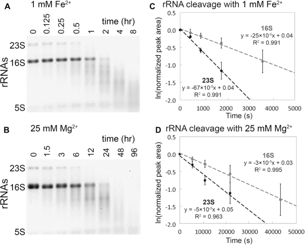
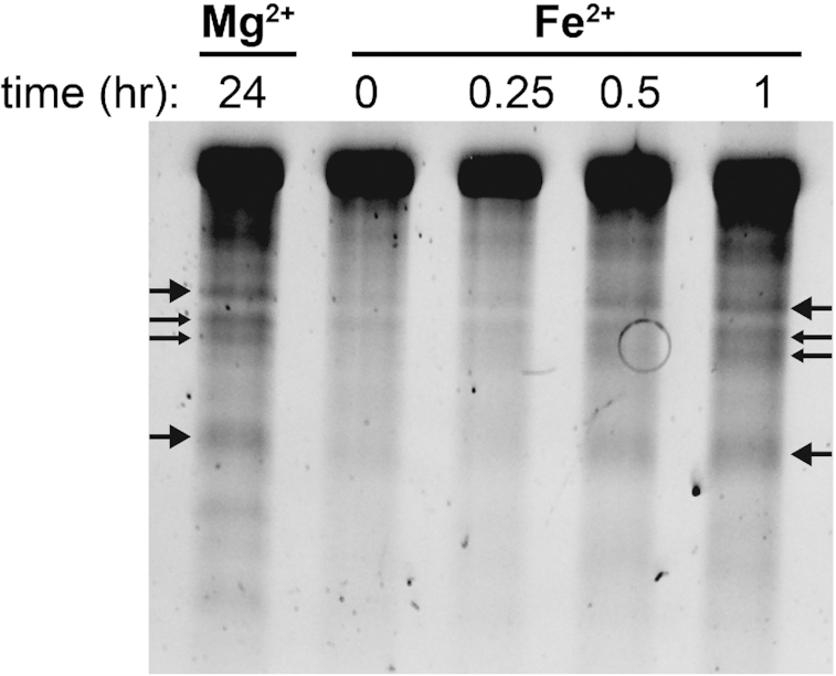
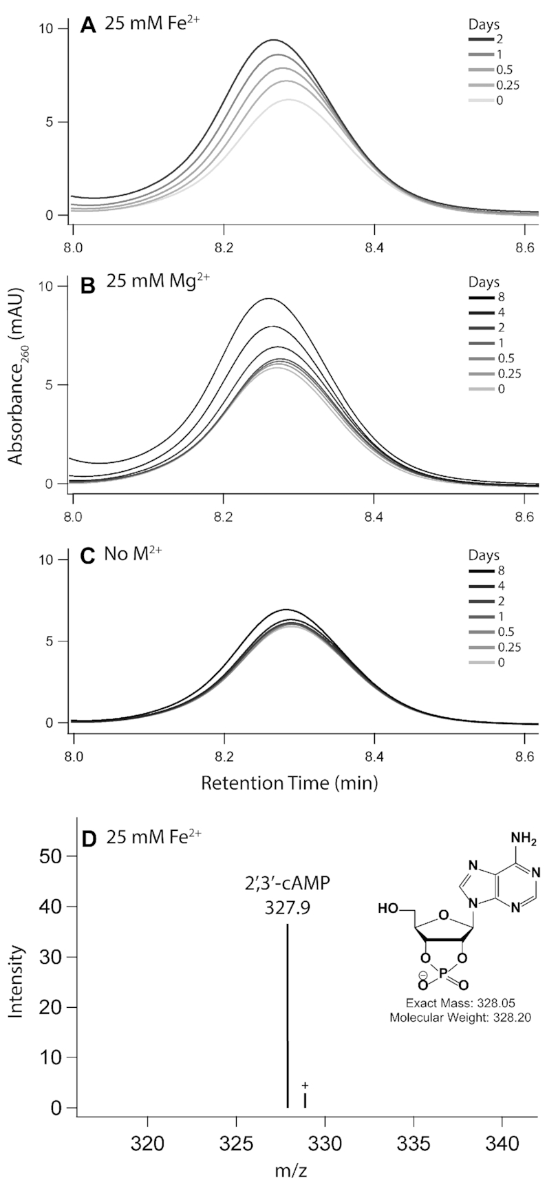
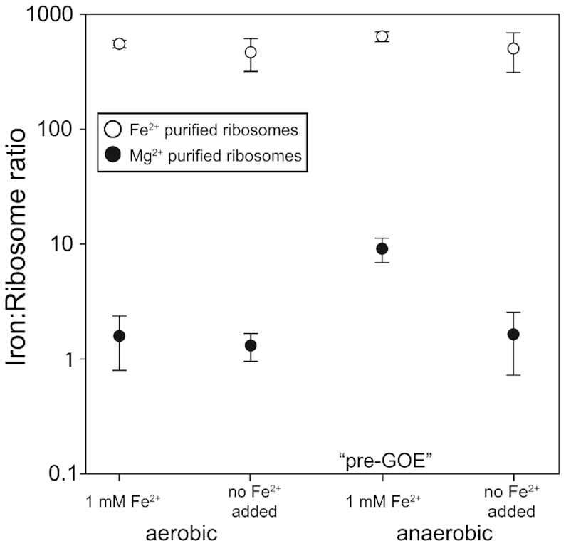
Divalent metal cations are essential to the structure and function of the ribosome. Previous characterizations of the ribosome performed under standard laboratory conditions have implicated Mg2+ as a primary mediator of ribosomal structure and function. Possible contributions of Fe2+ as a ribosomal cofactor have been largely overlooked, despite the ribosome's early evolution in a high Fe2+ environment, and the continued use of Fe2+ by obligate anaerobes inhabiting high Fe2+ niches. Here, we show that (i) Fe2+ cleaves RNA by in-line cleavage, a non-oxidative mechanism that has not previously been shown experimentally for this metal, (ii) the first-order in-line rate constant with respect to divalent cations is >200 times greater with Fe2+ than with Mg2+, (iii) functional ribosomes are associated with Fe2+ after purification from cells grown under low O2 and high Fe2+ and (iv) a small fraction of Fe2+ that is associated with the ribosome is not exchangeable with surrounding divalent cations, presumably because those ions are tightly coordinated by rRNA and deeply buried in the ribosome. In total, these results expand the ancient role of iron in biochemistry and highlight a possible new mechanism of iron toxicity.


Figures

Figure 1.
Figure 1.
In-line cleavage of rRNA in anoxia. In-line cleavage of purified rRNAs with (A) 1 mM Fe2+ (0–8 h) and (B) 25 mM Mg2+ (0–96 h). Reactions were conducted in an anoxic Coy chamber at 37°C in the presence of the hydroxyl radical quencher glycerol (5% v/v) and were analyzed by 1% agarose gel electrophoresis. Pseudo first-order rate plots were extracted from 23S and 16S rRNA band intensities for (C) 1 mM Fe2+ and (D) 25 mM Mg2+. The Mg2+ time axis is 10 times greater than the Fe2+ time axis. Error bars represent the SEM (standard error of the mean; n = 3).
Figure 2.
Figure 2.
In-line cleavage banding patterns are the same for rRNA cleavage with Mg2+ and anoxic Fe2+. Several primary cleavage bands of a-rRNA (40) are indicated by arrows. This gel is 6% polyacrylamide, 8 M urea showing in-line cleavage mediated by 1 mM Mg2+ or 1 mM anoxic Fe2+ at 37°C for varying amounts of time. Reactions were run in 20 mM Tris–HEPES (pH 7.2).
Figure 3.
Figure 3.
2′,3′-cAMP is formed upon incubation of ApA with Fe2+ or Mg2+. HPLC chromatograms show the accumulation of 2′,3′-cAMP, a direct product of in-line cleavage, upon incubation of ApA with either (A) 25 mM Fe2+, (B) 25 mM Mg2+ or (C) no metal as the negative control. Panel (D) shows identification of the 2′,3′-cyclic adenosine monophosphate by LC–MS of ApA incubated with 25 mM Fe2+ for 2 days. Labeled species correspond to [M−H] ions. Reactions were incubated anoxically at 37°C in the presence of 5% (v/v) glycerol.
Figure 4.
Figure 4.
Iron content (mol Fe mol−1 ribosome) of purified ribosomes. E. coli were grown aerobically or anaerobically at 1 mM Fe2+ or ambient Fe2+ (6–9 μM, no Fe added), and purified in buffers containing either 3 mM Mg2+ (black circles) or 1 mM Fe2+ (white circles). Error bars represent the SEM (n = 3).
Figure 5.
Figure 5.
Agarose gels (1%) showing rRNA from ribosomes purified in (A) 3 mM Mg2+ and (B) 1 mM Fe2+. The banding pattern suggests that rRNA is less degraded in ribosomes purified with 3 mM Mg2+ than in ribosomes purified with 1 mM Fe2+.

References

    1. Bernier C.R., Petrov A.S., Kovacs N.A., Penev P.I., Williams L.D.. Translation: the universal structural core of life. Mol. Biol. Evol. 2018; 35:2065–2076. - PMC - PubMed
    1. Melnikov S., Ben-Shem A., Garreau de Loubresse N., Jenner L., Yusupova G., Yusupov M.. One core, two shells: bacterial and eukaryotic ribosomes. Nat. Struct. Mol. Biol. 2012; 19:560–567. - PubMed
    1. Woese C.R. Translation: in retrospect and prospect. RNA. 2001; 7:1055–1067. - PMC - PubMed
    1. Noller H.F., Kop J., Wheaton V., Brosius J., Gutell R.R., Kopylov A.M., Dohme F., Herr W., Stahl D.A., Gupta R.. Secondary structure model for 23S ribosomal RNA. Nucleic Acids Res. 1981; 9:6167–6189. - PMC - PubMed
    1. Petrov A.S., Bernier C.R., Hsiao C., Norris A.M., Kovacs N.A., Waterbury C.C., Stepanov V.G., Harvey S.C., Fox G.E., Wartell R.M. et al. .. Evolution of the ribosome at atomic resolution. Proc. Natl. Acad. Sci. U.S.A. 2014; 111:10251–10256. - PMC - PubMed

Publication types