Review
. 2020 Sep:198:108137.
doi: 10.1016/j.exer.2020.108137. Epub 2020 Jul 11.

Composition, structure and function of the corneal stroma

Affiliations
Review

Composition, structure and function of the corneal stroma

Edgar M Espana et al. Exp Eye Res. 2020 Sep.

Abstract

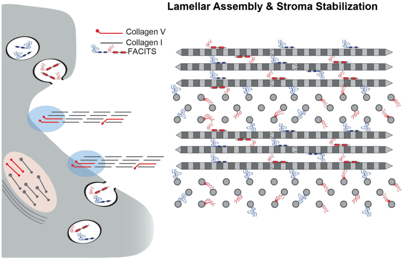
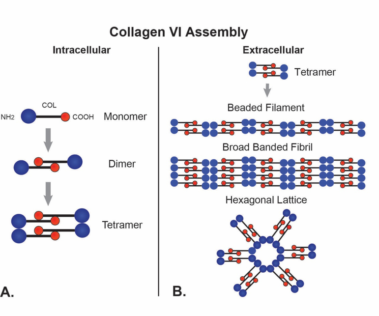
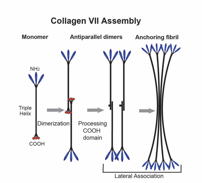
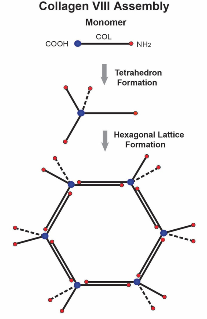
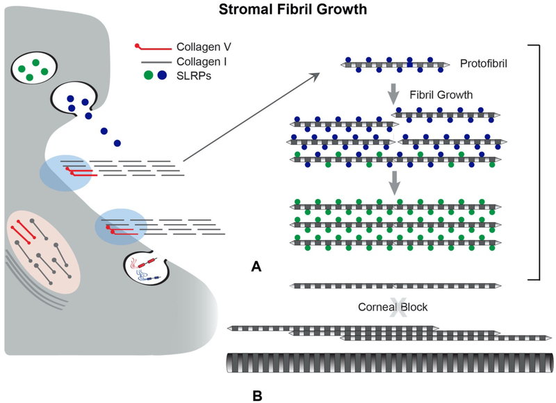
No other tissue in the body depends more on the composition and organization of the extracellular matrix (ECM) for normal structure and function than the corneal stroma. The precise arrangement and orientation of collagen fibrils, lamellae and keratocytes that occurs during development and is needed in adults to maintain stromal function is dependent on the regulated interaction of multiple ECM components that contribute to attain the unique properties of the cornea: transparency, shape, mechanical strength, and avascularity. This review summarizes the contribution of different ECM components, their structure, regulation and function in modulating the properties of the corneal stroma. Fibril forming collagens (I, III, V), fibril associated collagens with interrupted triple helices (XII and XIV), network forming collagens (IV, VI and VIII) as well as small leucine-rich proteoglycans (SLRP) expressed in the stroma: decorin, biglycan, lumican, keratocan, and fibromodulin are some of the ECM components reviewed in this manuscript. There are spatial and temporal differences in the expression of these ECM components, as well as interactions among them that contribute to stromal function. Unique regions within the stroma like Bowman's layer and Descemet's layer are discussed. To define the complexity of corneal stroma composition and structure as well as the relationship to function is a daunting task. Our knowledge is expanding, and we expect that this review provides a comprehensive overview of current knowledge, definition of gaps and suggests future research directions.

Keywords: Collagen fibril; Collagens; Composition; Cornea; Proteoglycans; Stroma; Structure.


Figures

Fig. 1.
Fig. 1.. Fibril-Forming Collagens: Fibrils.
Collagen I is the major stromal protein. This fibril-forming collagen is synthesized as procollagen. Procollagens have a central collagen (COL) domain with flanking N-and C-terminal propeptides. Extracellularly, the propeptides are processed and the resulting collagen molecules assemble to form striated fibrils. The collagen I molecule is approximately 300 nm in length and 1.5 nm in diameter. During fibril assembly, the collagen molecules are staggered N to C and this staggered pattern of collagen molecules gives rise to a 67nm repeat. An electron micrograph of a negative stained fibril is shown at the bottom of the panel. This fibril has the characteristic alternating light/dark pattern representing the gap (dark) and overlap (light) regions of the fibril.
Fig. 2.
Fig. 2.. Corneal Stroma Heterotypic Fibrils.
Corneal stromal collagen fibrils are heterotypic, co-assembled from quantitatively a major fibril forming collagen, e.g., collagen I and regulatory fibril-forming collagen, e.g., collagen V. Regulatory fibril-forming collagens have a partially processed N-terminal propeptide, retaining a non-collagenous domain that must be in/on the gap region/fibril surface. This is the major regulatory domain.
Fig. 3.
Fig. 3.. Heterotypic Collagen I/V Interactions Regulate Initial Fibril Assembly.
Stromal fibril assembly involves a sequence of regulatory interactions. (A) Initially, collagen V interacts with collagen I to nucleate collagen assembly into fibrils at the keratocyte surface. This results in the regulated assembly of immature, small diameter, short fibrils termed protofibrils. This initial step in fibril assembly is cell-directed involving interactions with organizers at the keratocyte surface, e.g., integrins and syndecans either directly or through intermediate interactions, e.g. fibronectin (indicated by shaded blue region). This permits kertocyte control over the initial assembly steps and allows for positioning of newly assembled fibrils into the stromal matrix. (B) In the absence of collagen V regulation of collagen I assembly and positioning of assembled fibrils is lost. This results in formation of fewer, larger and heterogenoeus fibril diameters, structurally abberent fibrils as well as disrupted fibril organization. This unregulated assembly is not consistent with stromal transparency.
Fig. 4.
Fig. 4.. FACIT Collagens.
Collagens XII and XIV are fibril-associated in the stroma. Their domain structures are illustrated. All FACITs have alternativly spliced variants and collagen XII can have glycosaminoglycan chains covalently attached. The FACIT collagens have 2–3 collagen (COL) domains and 3–4 non-collagenous (NC) domains. Characteristic of this collagen type is a large N-terminal NC domain that projects into the inter-fibrillar space. The FACIT collagens all associate with the surface of collagen fibrils and this is illustrated, including N-truncated isoforms due to alternative splicing in collagen XII. Collagen XII is capable of non-fibrillar interactions (not shown), it is not known if this is true for collagen XIV.
Fig. 5.
Fig. 5.. FACIT Regulation of Stromal Hierarchal Assembly.
FACIT collagens bind to fibril surfaces. In addition, they can interact with cells and a number of other matrix components. These properties are required for integration of different stromal components. FACIT collagens XII and XIV are associated with stromal fibrils. These interactions, along with SLRPs (see Fig. 9) can stabilize fibril diameter and spacing. In the absence of collagen XII, stromal lamellae fail to develop properly and there is a general disorganization of lammelar and stromal architecture. This indicates a critical role in integration of the fibrillar components necessary for stable lamellae formation. There is also evidence that collagen XII is enriched at cell and basement membrane interfaces (not shown) and may facilitate cell-matrix integration.
Fig. 6.
Fig. 6.. Collagen VI.
Collagen VI forms networks of beaded filaments in the corneal stroma. (A) Collagen VI monomers have a C-terminal non-collagenous (NC) domain, a central triple helical domain and an N-terminal NC domain. Intracellularly, monomers assemble N-C to form dimmers. Tetramers are assembled from two dimmers aligned in register. (B) The tetramers are secreted, and extracellularly they form the building blocks of 3 different collagen VI Assemblies. These include beaded filaments, broad banded fibrils and hexagonal lattices form via end-to-end interactions of tetramers and varying degrees of lateral association.
Fig. 7.
Fig. 7.. Collagen VII.
Collagen VII forms a network of anchoring fibrils that adhere to the epithelial basement membrane and form loops entrapping the collagen fibrils in Bowman’s layer. This integrates the epithelial basement membrane with the underlying stroma. Collagen VII has a long central triple-helical collagenous (COL) domain containing numerous interruptions conferring flexibility to the domain. The COL domain is flanked by non-collagenous N- (NC-1) and C-terminal (NC-2) domains. Two monomers interact to form an anti-parallel dimer with a central C-terminal overlap and the NC-1 domains pointing out. Processing occurs, with a cleavage of the NC-2 propeptide and covalent stabilization of the dimer. At this point, a non-staggered lateral association of dimers occurs that produces anchoring fibrils.
Fig. 8.
Fig. 8.. Collagen VIII Forms Hexagonal Lattices In Descemet’s. Membrane.
Collagen VIII is a short chain collagen with a central collagenous (COL) domain and flanking N- and C-terminal non-collagenous (NC) domains. The C-terminal NC domains of 4 collagen VIII molecules interact to form tetrahedrons. Tetrahedrons assemble further to form hexagonal lattices. A planar hexagonal lattice is illustrated. In Descemet’s membrane continued assembly, involving interactions of the N-terminal NC domains or anti-parallel interactions involving both helical and terminal domains (not shown) generates the layered hexagonal lattice characteristic of Descemet’s membrane (not shown).
Fig 9.
Fig 9.. SLRPs Regulate Lateral Fibril Growth.
SLRPs regulate linear and lateral stromal collagen fibril growth by binding to fibril surfaces. (A) Newly assembled stromal fibrils (protofibrils) are deposited into the matrix where they are stabilized via interactions with SLRPs. After deposition into the stromal matrix, protofibrils mature by a process of fibril growth. Bound SLRPs mediate and controlled fibrillar interactions resulting in coordinated growth of mature stromal fibrils. In the stroma there is a a tissue-specific regulation of lateral fibril growth. Diameter and packing are rigidly regulated for corneal transparency. (B) In most tissues there is a robust lateral growth resulting in heterogeneous populations of large diameter fibrils this is blocked in the stroma. Changes in fibril stabilization necessary for growth can result from processing, turnover, and/or displacement of SLRPs. SLRPs affect fibril diameter and spacing in the corneal stroma. When SLRPs are absent, as in null mice or in gene mutations in human patients, fibril structure, organization and spacing are impacted, compromising transparency. Also, the block to lateral fibril growth is lost in the stroma when SLPR expression is altered.

References

    1. Agah A, Kyriakides TR, Lawler J, Bornstein P, 2002. The lack of thrombospondin-1 (TSP1) dictates the course of wound healing in double-TSP1/TSP2-null mice. Am J Pathol 161, 831–839. - PMC - PubMed
    1. Akama TO, Nishida K, Nakayama J, Watanabe H, Ozaki K, Nakamura T, Dota A, Kawasaki S, Inoue Y, Maeda N, Yamamoto S, Fujiwara T, Thonar EJ, Shimomura Y, Kinoshita S, Tanigami A, Fukuda MN, 2000. Macular corneal dystrophy type I and type II are caused by distinct mutations in a new sulphotransferase gene. Nat Genet 26, 237–241. - PubMed
    1. Ansorge HL, Meng X, Zhang G, Veit G, Sun M, Klement JF, Beason DP, Soslowsky LJ, Koch M, Birk DE, 2009. Type XIV Collagen Regulates Fibrillogenesis: PREMATURE COLLAGEN FIBRIL GROWTH AND TISSUE DYSFUNCTION IN NULL MICE. J Biol Chem 284, 8427–8438. - PMC - PubMed
    1. Arai K, Nagashima Y, Takemoto T, Nishiyama T, 2008. Mechanical strain increases expression of type XII collagen in murine osteoblastic MC3T3-E1 cells. Cell Struct Funct 33, 203–210. - PubMed
    1. Armstrong DJ, Hiscott P, Batterbury M, Kaye S, 2003. Keratocyte matrix interactions and thrombospondin 2. Mol Vis 9, 74–79. - PubMed