Review
. 2020 Oct:94:102903.
doi: 10.1016/j.dnarep.2020.102903. Epub 2020 Jun 29.

Structural biology of DNA abasic site protection by SRAP proteins

Affiliations
Review

Structural biology of DNA abasic site protection by SRAP proteins

Katherine M Amidon et al. DNA Repair (Amst). 2020 Oct.

Abstract

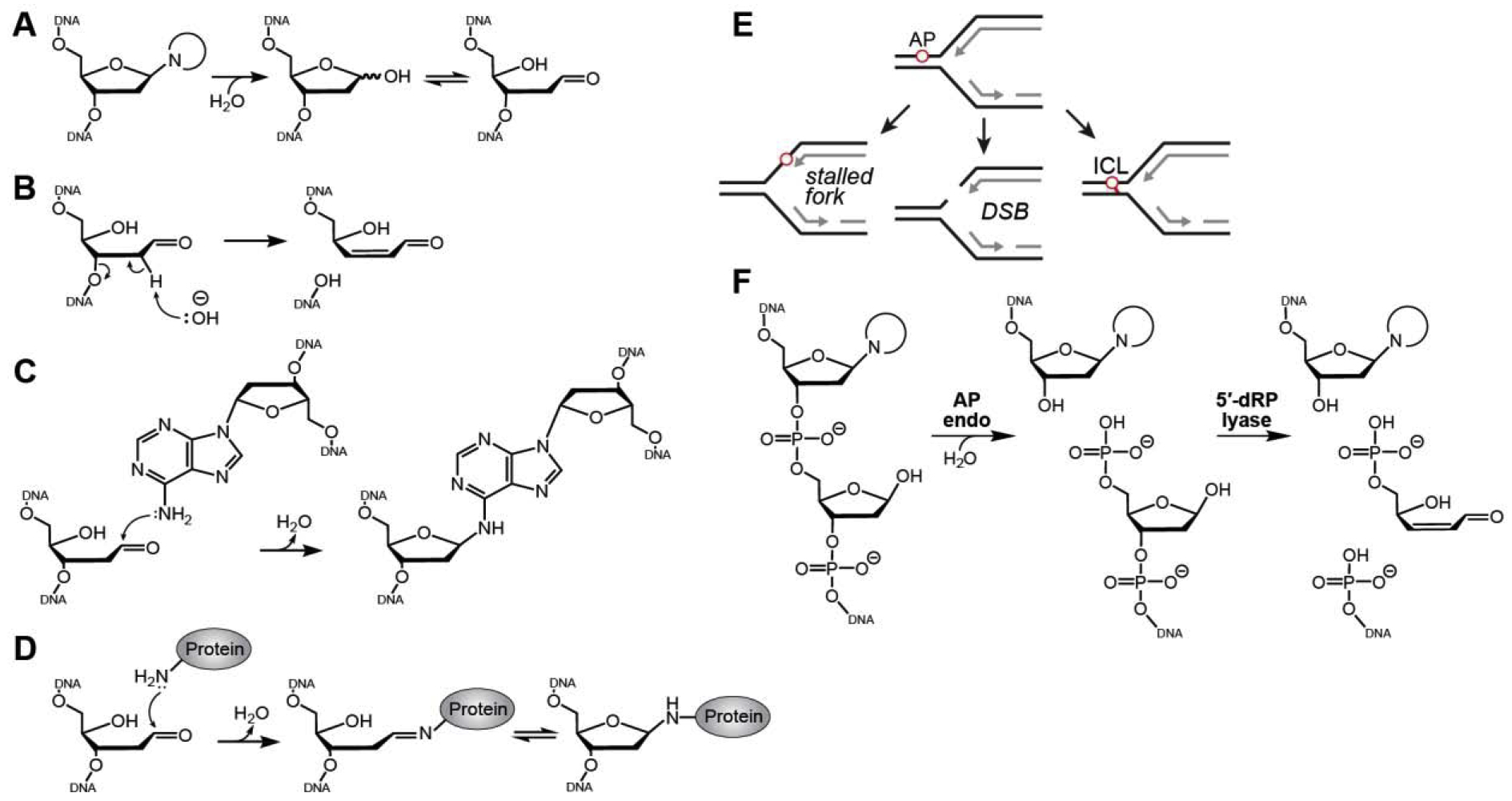
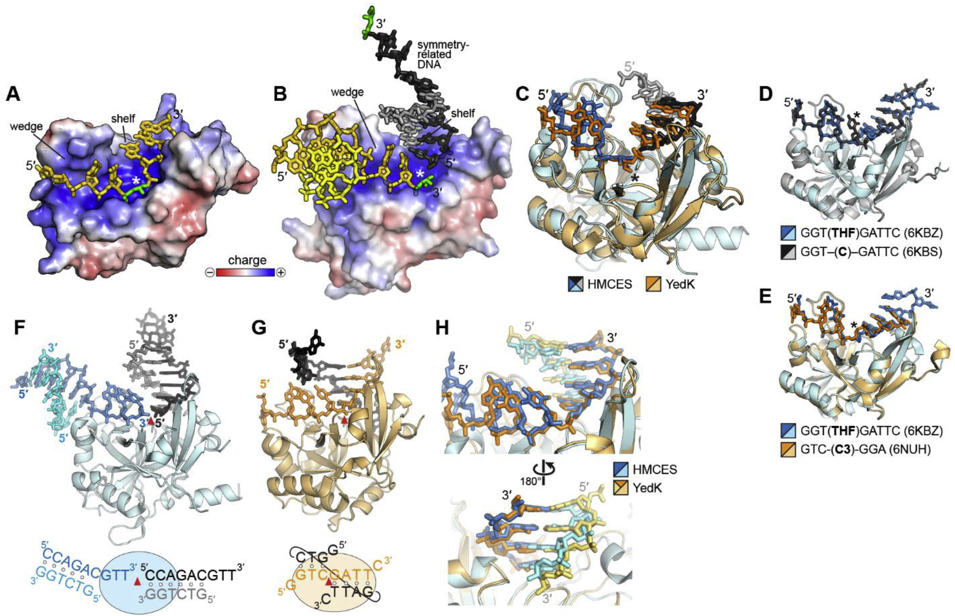
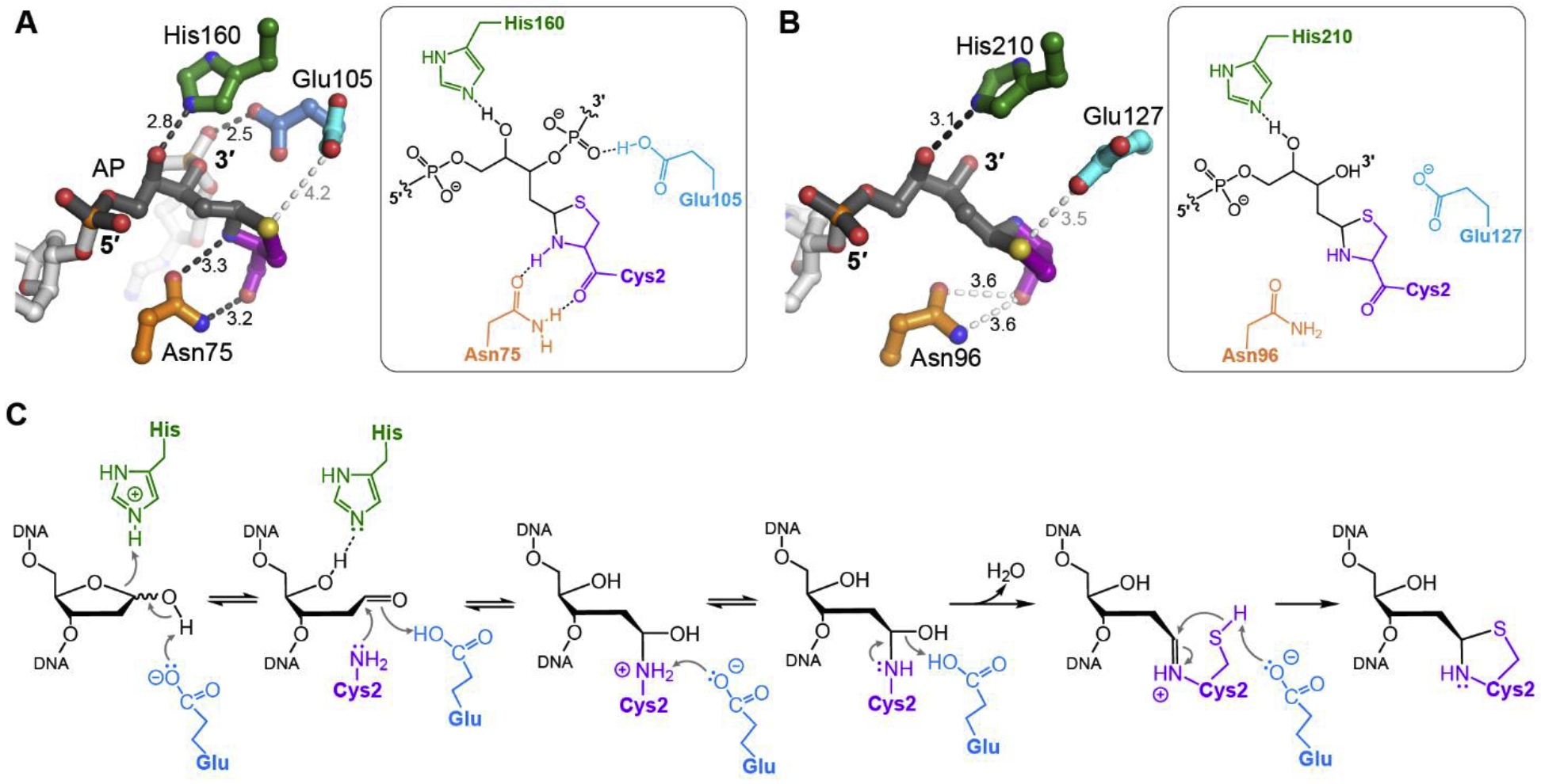
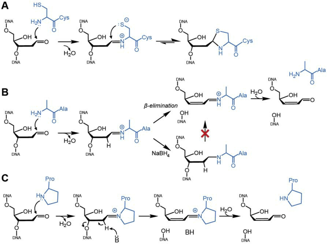
Abasic (AP) sites are one of the most frequently occurring types of DNA damage. They lead to DNA strand breaks, interstrand DNA crosslinks, and block transcription and replication. Mutagenicity of AP sites arises from translesion synthesis (TLS) by error-prone bypass polymerases. Recently, a new cellular response to AP sites was discovered, in which the protein HMCES (5-hydroxymethlycytosine (5hmC) binding, embryonic stem cell-specific) forms a stable, covalent DNA-protein crosslink (DPC) to AP sites at stalled replication forks. The stability of the HMCES-DPC prevents strand cleavage by endonucleases and mutagenic bypass by TLS polymerases. Crosslinking is carried out by a unique SRAP (SOS Response Associated Peptidase) domain conserved across all domains of life. Here, we review the collection of recently reported SRAP crystal structures from human HMCES and E. coli YedK, which provide a unified basis for SRAP specificity and a putative chemical mechanism of AP site crosslinking. We discuss the structural and chemical basis for the stability of the SRAP DPC and how it differs from covalent protein-DNA intermediates in DNA lyase catalysis of strand scission.

Keywords: Abasic site; DNA lyase; DNA-protein crosslink; HMCES; SRAP; Thiazolidine.


Conflict of interest statement

DECLARATION OF COMPETING INTEREST

There are no conflicts of interest to declare.

Figures

Fig. 1.
Fig. 1.. Consequences of abasic sites.
A. AP sites arise from enzymatic and spontaneous hydrolysis of the N-glycosidic bond and exist in either furanose or aldehyde forms. B. Base-catalyzed β-elimination of an AP site generates a strand break. C,D. Formation of an ICL (C) and a DPC (D) by nucleophilic attack of AP site C1ʹ by primary amines in DNA or proteins . E. Consequences of AP sites in the template strand during DNA replication. F. Incision of DNA by AP endonuclease and DNA lyases.
Fig. 2.
Fig. 2.. SRAP-DNA structure.
A,B. Electrostatic surface potential of (A) YedK-DPC (PDB ID 6NUA and (B) HMCES-SRAP DPC (PDB ID 6OE7) structures. The AP sites are green, and the symmetry-related DNA molecule in HMCES is shown in black and grey. The white asterisk denotes the position of the active site. C. Superposition of YedK-DPC (PDB ID 6NUA) and HMCES-SRAP DPC (PDB ID 6OE7). The DPC is marked with an asterisk. The ends of the DNA in the HMCES structure have been removed for clarity. D,E. Superposition of YedK bound noncovalently to ssDNA containing (D) a cytosine or an abasic site analog, and (E) two different abasic site analogs. The position of the nucleotide at the active site is marked with an asterisk. F,G. Similarity of dsDNA bound to (F) HMCES-SRAP (PDB ID 6OEB) and (G) YedK (PDB ID 6KBS). The asymmetric unit is colored blue or orange, and symmetry related DNA is shown in black and grey. The red triangle marks the position of the active site. In the schematic at the bottom, base pairs are denoted by open circles. H. Superposition of the two structures in panels F and G. DNA is colored blue/cyan in HMCES and orange/gold in YedK.
Fig. 3.
Fig. 3.. SRAP active site and mechanism of crosslinking.
A,B. Atomic details of the thiazolidine DPC in (A) YedK (PDB ID 6NUA) and (B) HMCES-SRAP (PDB ID 6OE7). DNA is greyscale with AP site dark grey, and SRAP is colored by amino acid. Interatomic distances (Å) are labeled, with hydrogen bonds indicated by dark dashes and close contacts with light dashes. C. Proposed catalytic mechanism of crosslinking.
Fig. 4.
Fig. 4.. Comparison of DPCs formed by SRAP and AP lyases.
A. Wild-type SRAP forms a thiazolidine DPC via a Schiff base intermediate. B. The Schiff base formed by a SRAP C2A mutant is prone to β-elimination and can be reduced to a stable DPC via borohydride treatment. C. Catalysis of DNA lyase activity by bifunctional DNA glycosylases.

References

    1. Nakamura J, Walker VE, Upton PB, Chiang S-Y, Kow YW and Swenberg JA (1998) Highly sensitive apurinic/apyrimidinic site assay can detect spontaneous and chemically induced depurination under physiological conditions. Cancer research, 58, 222–225. - PubMed
    1. Nakamura J and Swenberg JA (1999) Endogenous apurinic/apyrimidinic sites in genomic DNA of mammalian tissues. Cancer research, 59, 2522–2526. - PubMed
    1. Lindahl T (1993) Instability and decay of the primary structure of DNA. nature, 362, 709–715. - PubMed
    1. Barnes DE and Lindahl T (2004) Repair and Genetic Consequences of Endogenous DNA Base Damage in Mammalian Cells. Annual Review of Genetics, 38, 445–476. - PubMed
    1. Cadet J and Wagner JR (2013) DNA base damage by reactive oxygen species, oxidizing agents, and UV radiation. Cold Spring Harb Perspect Biol, 5. - PMC - PubMed

Publication types

LinkOut - more resources