. 2020 Jul 10;13(14):3082.
doi: 10.3390/ma13143082.

Electrochemical Analysis and In Vitro Assay of Mg-0.5Ca-xY Biodegradable Alloys

Affiliations

Electrochemical Analysis and In Vitro Assay of Mg-0.5Ca-xY Biodegradable Alloys

Bogdan Istrate et al. Materials (Basel). .

Abstract

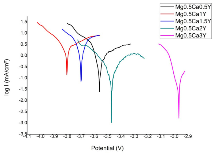
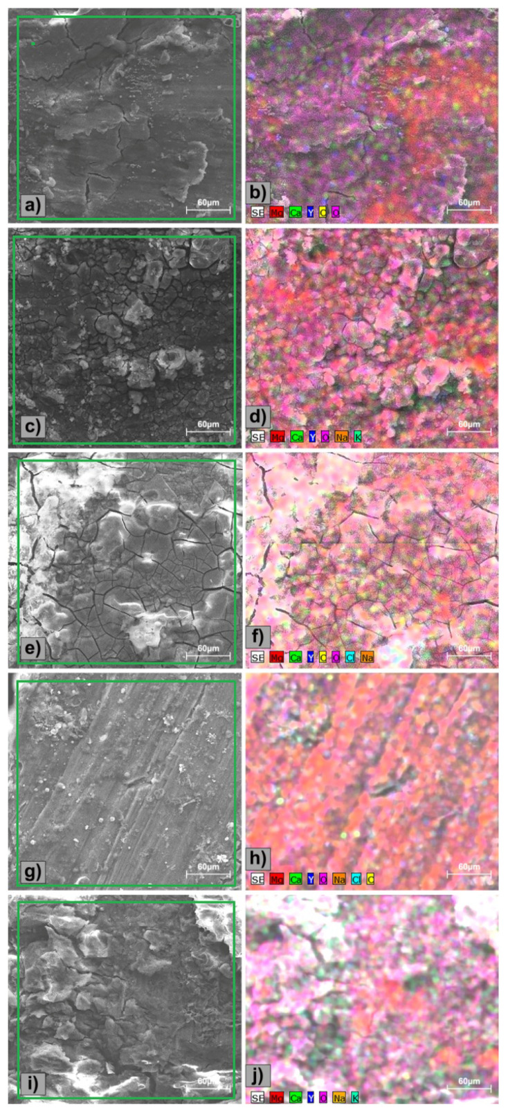
In recent years, biodegradable Mg-based materials have been increasingly studied to be used in the medical industry and beyond. A way to improve biodegradability rate in sync with the healing process of the natural human bone is to alloy Mg with other biocompatible elements. The aim of this research was to improve biodegradability rate and biocompatibility of Mg-0.5Ca alloy through addition of Y in 0.5/1.0/1.5/2.0/3.0wt.%. To characterize the chemical composition and microstructure of experimental Mg alloys, scanning electron microscopy (SEM), energy-dispersive spectroscopy (EDS), light microscopy (LM), and X-ray diffraction (XRD) were used. The linear polarization resistance (LPR) method was used to calculate corrosion rate as a measure of biodegradability rate. The cytocompatibility was evaluated by MTT assay (3-(4,5-dimethylthiazole-2-yl)-2,5-diphenyltetrazolium bromide) and fluorescence microscopy. Depending on chemical composition, the dendritic α-Mg solid solution, as well as lamellar Mg2Ca and Mg24Y5 intermetallic compounds were found. The lower biodegradability rates were found for Mg-0.5Ca-2.0Y and Mg-0.5Ca-3.0Y which have correlated with values of cell viability. The addition of 2-3 wt.%Y in the Mg-0.5Ca alloy improved both the biodegradability rate and cytocompatibility behavior.

Keywords: Mg-Ca-Y alloys; electrochemical evaluation; in vitro test; microstructure.


Conflict of interest statement

The authors declare no conflict of interest.

Figures

Figure 1
Figure 1
Light microscopy analysis of the Mg-0.5Ca-xY: (a) Mg-0.5Ca-0.5Y; (b) Mg-0.5Ca-1.0Y [57]; (c) Mg-0.5Ca-1.5Y; (d) Mg-0.5Ca-2.0Y [57]; (e) Mg-0.5Ca-3.0Y [57].
Figure 2
Figure 2
Surface SEM images of the Mg-0.5Ca-xY: (a) Mg-0.5Ca-0.5Y; (b) Mg-0.5Ca-1.0Y [57]; (c) Mg-0.5Ca-1.5Y; (d) Mg-0.5Ca-2.0Y [57]; (e) Mg-0.5Ca-3.0Y [57].
Figure 3
Figure 3
XRD analysis of Mg-0.5Ca-xY experimental alloys [57].
Figure 4
Figure 4
Tafel diagrams of Mg-0.5Ca-xY-based experimental alloys.
Figure 5
Figure 5
SEM images of experimental surfaces after electrochemical tests: (a,b) Mg-0.5Ca-0.5Y; (c,d) Mg-0.5Ca-1.0Y; (e,f) Mg-0.5Ca-1.5Y; (g,h) Mg-0.5Ca-2.0Y; and (i,j) Mg-0.5Ca-3.0Y.
Figure 6
Figure 6
Distribution of the identified elements on the surface of the Mg-0.5Ca-xY alloys after electrochemical tests: (a,b) Mg-0.5Ca-0.5Y; (c,d) Mg-0.5Ca-1.0Y; (e,f) Mg-0.5Ca-1.5Y; (g,h) Mg-0.5Ca-2.0Y; and (i,j) Mg-0.5Ca-3.0Y.
Figure 7
Figure 7
Cell viability profile (%) evaluated by 3-(4,5-dimethylthiazole-2-yl)- 2,5-diphenyltetrazolium bromide - (MTT)-assay: Effect of Mg-0.5Ca-xY experimental alloys on cell viability after 1, 3, and 5 days of co-incubation. Data expressed as percent related to the negative control. (˄; o; ●; ˅; □) No significant differences on cell viability (p > 0.05); see details in the text.
Figure 8
Figure 8
Variation of the pH in the DMEM-F12 complete media (Dulbecco’s modified Eagle medium/Nutrient F-12 Ham) during co-incubation with the studied Mg-0.5Ca-xY alloys samples (at 37 °C, 5% CO2), over a period of 5 days.
Figure 9
Figure 9
Fluorescence microscopy images of fibroblastic cells’ morphology after 1 and 5 days of coincubation with the experimental alloys. Viable cells stained in green because of calcein fluorescent dye presence inside the cells. Bar: 200 μm.

References

    1. Schumann P., Lindhorst D., Wagner M., Schramm A., Gellrich N.C., Rucker M. Perspectives on ResorbableOsteosynthesis Materials in Cranio maxilla facial Surgery. Pathobiology. 2013;80:211–217. doi: 10.1159/000348328. - DOI - PubMed
    1. Rahim M.I., Ullah S., Mueller P. Advances and Challenges of Biodegradable Implant Materials with a Focus on Magnesium-Alloys and Bacterial Infections. Metals. 2018;8:532. doi: 10.3390/met8070532. - DOI
    1. Banerjee P.C., Al-Saadi S., Choudhary L., Harandi S.E., Singh R. Magnesium Implants: Prospects and Challenges. Materials. 2019;12:136. doi: 10.3390/ma12010136. - DOI - PMC - PubMed
    1. Tune D. Body-Absorbable Osteosynthesis Devices. Clin. Mater. 1991;8:119–123. doi: 10.1016/0267-6605(91)90019-c. - DOI - PubMed
    1. Staiger M.P., Pietak A.M., Huadmai J., Dias G. Magnesium and its alloys as orthopedic biomaterials: A review. Biomaterials. 2006;27:1728–1734. doi: 10.1016/j.biomaterials.2005.10.003. - DOI - PubMed

LinkOut - more resources