. 2020 Jul 11;21(14):4896.
doi: 10.3390/ijms21144896.

The Soluble Adenylyl Cyclase Inhibitor LRE1 Prevents Hepatic Ischemia/Reperfusion Damage Through Improvement of Mitochondrial Function

Affiliations

The Soluble Adenylyl Cyclase Inhibitor LRE1 Prevents Hepatic Ischemia/Reperfusion Damage Through Improvement of Mitochondrial Function

João S Teodoro et al. Int J Mol Sci. .

Abstract

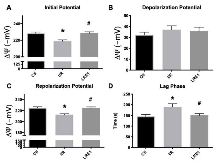
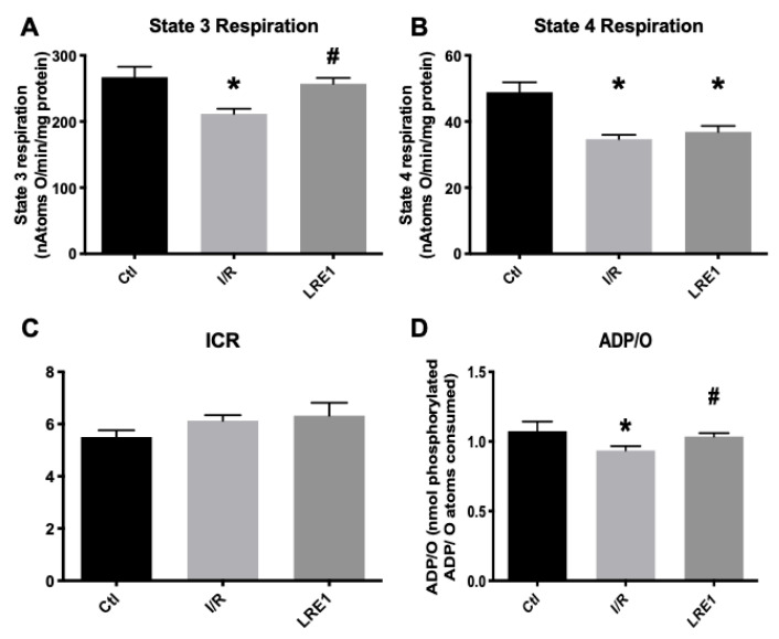
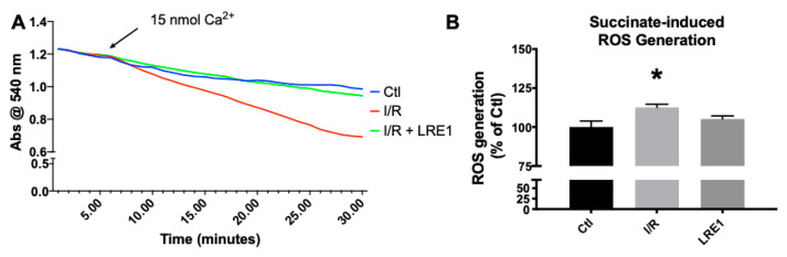
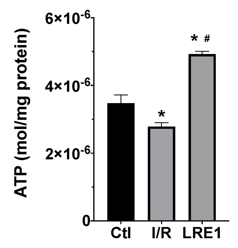
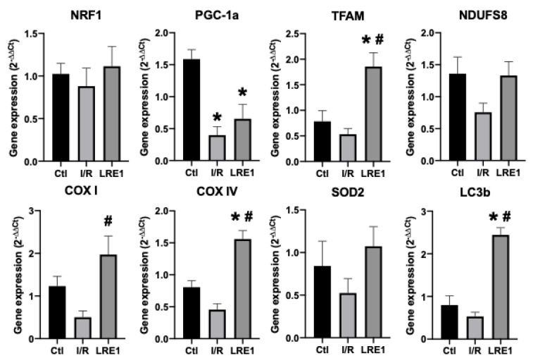
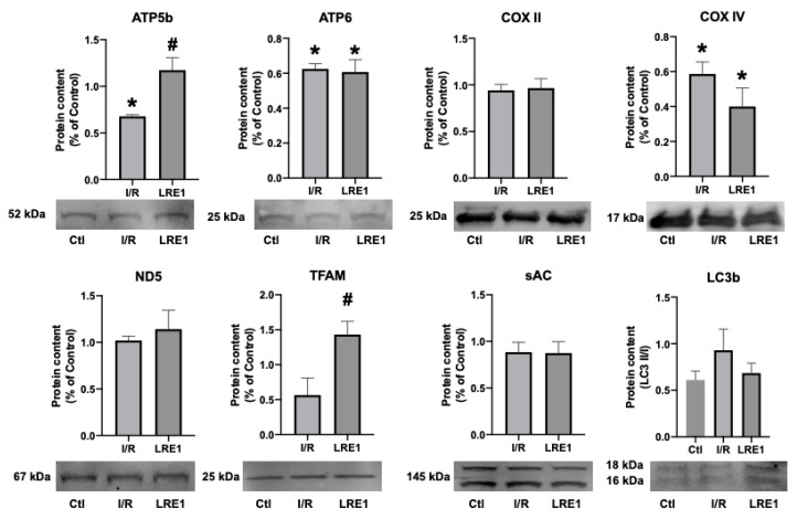
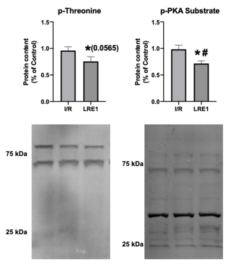
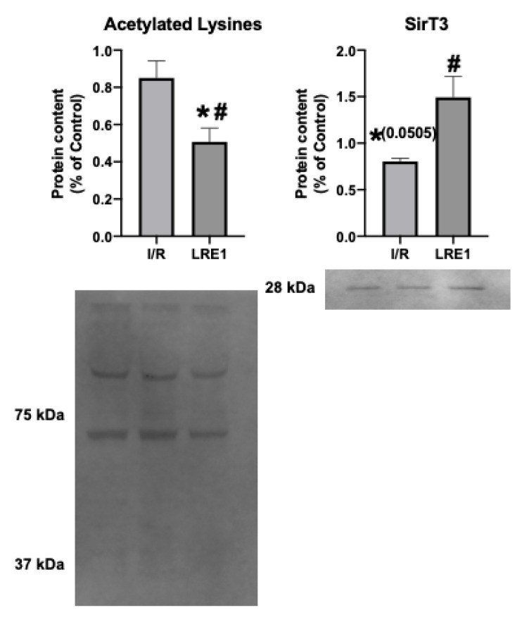
Hepatic ischemia/reperfusion (I/R) injury is a leading cause of organ dysfunction and failure in numerous pathological and surgical settings. At the core of this issue lies mitochondrial dysfunction. Hence, strategies that prime mitochondria towards damage resilience might prove applicable in a clinical setting. A promising approach has been to induce a mitohormetic response, removing less capable organelles, and replacing them with more competent ones, in preparation for an insult. Recently, a soluble form of adenylyl cyclase (sAC) has been shown to exist within mitochondria, the activation of which improved mitochondrial function. Here, we sought to understand if inhibiting mitochondrial sAC would elicit mitohormesis and protect the liver from I/R injury. Wistar male rats were pretreated with LRE1, a specific sAC inhibitor, prior to the induction of hepatic I/R injury, after which mitochondria were collected and their metabolic function was assessed. We find LRE1 to be an effective inducer of a mitohormetic response based on all parameters tested, a phenomenon that appears to require the activity of the NAD+-dependent sirtuin deacylase (SirT3) and the subsequent deacetylation of mitochondrial proteins. We conclude that LRE1 pretreatment leads to a mitohormetic response that protects mitochondrial function during I/R injury.

Keywords: LRE1; ischemia/reperfusion; liver; mitochondria; sirtuin 3; soluble adenylyl cyclase.


Conflict of interest statement

D.A.S. is a consultant to, inventor of patents licensed to, and, in some cases, board member and investor of MetroBiotech, CohBar, Life Biosciences and affiliates, InsideTracker, Vium, Zymo, EdenRoc Sciences and affiliates, Immetas, Segterra, Galilei Biosciences, and Iduna Therapeutics. He is also an inventor on patent applications licensed to Bayer Crops, Merck KGaA, and Elysium Health. For details see https://genetics.med.harvard.edu/sinclair. The other authors report no conflicts of interest.

Figures

Figure 1
Figure 1
Mitochondrial membrane initial potential (A), ADP-driven depolarization potential (B), repolarization potential (C) and lag phase time (D) as evaluated by tetraphenylphosphonium ion (TPP+) fluctuations and recorded with a TPP+-sensitive electrode. Data are means ± SEM of 4 independent experiments. * indicates a statistically significant difference vs. sham operated controls (Ctl); # indicates a statistically significant difference vs. ischemia/reperfusion (I/R).
Figure 2
Figure 2
Mitochondrial oxygen consumption parameters (ADP-driven State 3 respiration, A; repolarization respiration State 4, B) and resulting ratios (Respiratory Control Index, ICR, C and; ADP/O, D), as evaluated by O2 consumption and recorded with a Clark-type electrode. Data are means ± SEM of 4 independent experiments. * indicates a statistically significant difference vs. Ctl; # indicates a statistically significant difference vs. I/R.
Figure 3
Figure 3
Mitochondrial swelling recording (A) and reactive oxygen species (ROS) generation (B). Swelling was recorded by following absorbance of light at 540 nm after a Ca2+ challenge, while ROS was evaluated by following 2′,7′-dichlorodihydrofluorescein diacetate (H2DCFDA) fluorescence for 10 min. For panel A, lines indicate a representative recording of all of the assays, while panel B represents means ± SEM of 4 independent experiments. * indicates a statistically significant difference vs. Ctl.
Figure 4
Figure 4
Liver mitochondria ATP content as evaluated with a bioluminescent assay. Data are means ± SEM of 4 independent experiments. * indicates a statistically significant difference vs. Ctl; # indicates a statistically significant difference vs. I/R.
Figure 5
Figure 5
Liver gene expression evaluation by semi-qPCR. Data are means ± SEM of 4 independent experiments. NRF1, nuclear respiratory factor 1; PGC-1a, peroxisome proliferator-activated receptor gamma, coactivator 1 alpha; TFAM, mitochondrial transcription factor A; NDUFS8, NADH dehydrogenase iron–sulfur protein 8, mitochondrial; COX I, cytochrome c oxidase subunit I; COX IV, cytochrome c oxidase, subunit IV; SOD2, superoxide dismutase 2, mitochondrial; LC3b, microtubule-associated proteins 1A/1B light chain 3B. * indicates a statistically significant difference vs. Ctl; # indicates a statistically significant difference vs. I/R.
Figure 6
Figure 6
Hepatic mitochondrial protein content quantification by Western blot. Data are means ± SEM of 4 independent experiments. ATP5b, ATP synthase F1 subunit beta, mitochondrial; ATP6, ATP synthase FO subunit 6, mitochondrial; COX II, cytochrome c oxidase subunit II; COX IV, cytochrome c oxidase, subunit IV; ND5, NADH-ubiquinone oxidoreductase chain 5; TFAM, mitochondrial transcription factor A; sAC, soluble adenylyl cyclase; LC3b, microtubule-associated proteins 1A/1B light chain 3B. * indicates a statistically significant difference vs. Ctl; # indicates a statistically significant difference vs. I/R.
Figure 7
Figure 7
Hepatic mitochondrial protein phosphorylation content quantification by Western blot. Data are means ± SEM of 4 independent experiments. p-threonine, phosphorylated threonine residues; p-PKA substrate, phosphorylated PKA substrate. * indicates a statistically significant difference vs. Ctl; # indicates a statistically significant difference vs. I/R.
Figure 8
Figure 8
Hepatic mitochondrial protein acetylation status and NAD+-dependent deacetylase sirtuin 3 (SirT3) quantification by Western blot. Data are means ± SEM of 4 independent experiments. SirT3, Sirtuin 3. * indicates a statistically significant difference vs. Ctl; # indicates a statistically significant difference vs. I/R.

References

    1. Martins R.M., Pinto Rolo A., Soeiro Teodoro J., Furtado E., Caetano Oliveira R., Tralhão J.G., Marques Palmeira C. Addition of Berberine to Preservation Solution in an Animal Model of Ex Vivo Liver Transplant Preserves Mitochondrial Function and Bioenergetics from the Damage Induced by Ischemia/Reperfusion. Int. J. Mol. Sci. 2011;19:184–189. doi: 10.3390/ijms19010284. - DOI - PMC - PubMed
    1. Martins R.M., Teodoro J.S., Furtado E., Rolo A.P., Palmeira C.M., Tralhão J.G. Recent insights into mitochondrial targeting strategies in liver transplantation. Int. J. Med Sci. 2018;15:248. doi: 10.7150/ijms.22891. - DOI - PMC - PubMed
    1. Jaeschke H. Molecular mechanisms of hepatic ischemia-reperfusion injury and preconditioning. Am. J. Physiol. Gastrointest. Liver Physiol. 2003;284:G15–G26. doi: 10.1152/ajpgi.00342.2002. - DOI - PubMed
    1. Carden D.L., Granger D.N. Pathophysiology of ischaemia-reperfusion injury. J. Pathol. 2000;190:255–266. doi: 10.1002/(SICI)1096-9896(200002)190:3<255::AID-PATH526>3.0.CO;2-6. - DOI - PubMed
    1. Guan L.-Y. Mechanisms of hepatic ischemia-reperfusion injury and protective effects of nitric oxide. World J. Gastrointest. Surg. 2014;6:122. doi: 10.4240/wjgs.v6.i7.122. - DOI - PMC - PubMed

MeSH terms