Comparative Study
. 2020 Oct;184(2):1181-1193.
doi: 10.1104/pp.20.00294. Epub 2020 Jul 14.

Light-induced HY5 Functions as a Systemic Signal to Coordinate the Photoprotective Response to Light Fluctuation

Affiliations
Comparative Study

Light-induced HY5 Functions as a Systemic Signal to Coordinate the Photoprotective Response to Light Fluctuation

Xiaochun Jiang et al. Plant Physiol. 2020 Oct.

Abstract

Optimizing the photoprotection of different leaves as a whole is important for plants to adapt to fluctuations in ambient light conditions. However, the molecular basis of this leaf-to-leaf communication is poorly understood. Here, we used a range of techniques, including grafting, chlorophyll fluorescence, revers transcription quantitative PCR, immunoblotting, chromatin immunoprecipitation, and electrophoretic mobility shift assays, to explore the complexities of leaf-to-leaf light signal transmission and activation of the photoprotective response to light fluctuation in tomato (Solanum lycopersicum). We established that light perception in the top leaves attenuated the photoinhibition of both PSII and PSI by triggering photoprotection pathways in the bottom leaves. Local light promoted the accumulation and movement of LONG HYPOCOTYL5 from the sunlit local leaves to the systemic leaves, priming the photoprotective response of the latter to light fluctuation. By directly activating the transcription of PROTON GRADIENT REGULATION5 and VIOLAXANTHIN DE-EPOXIDASE, LONG HYPOCOTYL5 induced cyclic electron flow, the xanthophyll cycle, and energy-dependent quenching. Our findings reveal a systemic signaling pathway and provide insight into an elaborate regulatory network, demonstrating a pre-emptive advantage in terms of the activation of photoprotection and, hence, the ability to survive in a fluctuating light environment.


Figures

Figure 1.
Figure 1.
Light application systemically induces tolerance against photoinhibition in tomato. A, Fv/Fm and ΔP700max determined in the fourth leaves (systemic leaves) after exposure to HL stress for 0 to 30 min with (light) or without (dark) a preillumination onto the fifth to eighth leaves (local leaves) for 1 h. B, Fv/Fm and ΔP700max determined in the fourth leaves (systemic leaves) after exposure to HL stress for 20 min with a preillumination onto the fifth to eighth leaves for 0 to 60 min. Irradiance intensity is 1500 μmol m−2 s−1 for both the preillumination and HL stress treatments. During the preillumination treatment of the fifth to eighth leaves, the first to fourth leaves of the plant were kept in dark. The false-color code depicted above the image ranges from 0 (black) to 1.0 (purple), representing the degree of photoinhibition of PSII. The leaves were digitally extracted for comparison. Data are presented as the means of three biological replicates (± sd). Lowercse letters indicate significant differences (P < 0.05) according to Tukey’s test.
Figure 2.
Figure 2.
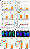
Light application triggers a photoprotective response in the systemic leaves of tomato. A, Gene expression of SlPGR5, SlVDE, and SlHY5. B, Half time of dark rereduction of P700+ (t1/2). C, Immunoblot analysis of VDE and PsbS proteins separated by SDS-PAGE. D, De-epoxidation state of the xanthophyll cycle in the fifth leaves (local) and the fourth leaves (systemic) in response to illumination onto the fifth to eighth leaves for 1 h. Plants in dark (dark) were used as controls. E, qE, and the kinetics of NPQ induction. Parameters for A–C and E were evaluated in the shaded fourth leaves (systemic leaves) after the fifth to eighth leaves were exposed to illumination at 1500 μmol m−2 s−1 for 0, 0.5, 1, or 2 h. During the illumination treatment, the first to fourth leaves of the plant were kept in dark. Data are presented as the means of three biological replicates (± sd). Lowercase letters indicate significant differences (P < 0.05) according to Tukey’s test.
Figure 3.
Figure 3.
Light application induces HY5 movement from local leaves to systemic leaves. A, Immunoblot analysis of HY5, VDE, and PsbS proteins separated by SDS-PAGE in the fifth leaves (local leaves) in response to illumination at 1500 μmol m−2 s−1 onto the fifth to eighth leaves for 0, 5, 10, 20, or 30 min. B, Immunoblot analysis of HY5 protein separated by SDS-PAGE in the fourth leaves (systemic leaves) after the fifth to eighth leaves were exposed to illumination at 1500 μmol m−2 s−1 for 0, 0.5, 1, or 2 h. C, Immunoblot analysis of the SlHY5-3HA fusion protein separated by SDS-PAGE in the fourth leaves after the exposure of the fifth to eighth leaves to illumination at 1500 μmol m−2 s−1 for 1 h. Wild type/wild type (WT) and HY5/wild type indicate young shoots with 4 leaves of wild-type plants and transgenic plants overexpressing HY5–3HA (HY5) grafted onto wild-type plants with 4 leaves, respectively. D, Immunoblot analysis of the SlHY5-3HA fusion protein separated by SDS-PAGE in the fifth leaves (local) and the fourth leaves (systemic) after the exposure of the fifth to eighth leaves to illumination at 1500 μmol m−2 s−1 for 0, 1, or 2 h. HY5/wild type plants were used for the determination of HY5 accumulation. During the illumination treatment of the fifth to eighth leaves, the first to fourth leaves of the plant were kept in dark.
Figure 4.
Figure 4.
HY5 expression abundance in the local leaves determines the sensitivity of systemic leaves to photoinhibition. Fv/Fm and ΔP700max were determined in the fourth leaves (systemic leaves) with a HL stress at 1500 μmol m−2 s−1 or without a HL stress (dark) for 20 min. Before that, the fifth to eighth leaves (local leaves) were exposed to a preillumination treatment for 1 h while the first to fourth leaves of the plant were kept in dark. hy5/wild type (WT), wild type/wild type, and HY5/wild type indicate plants with shoots of HY5-RNAi (hy5), wild-type, and HY5-overexpressing (HY5) plants grafted onto wild-type plants, respectively. The false-color code depicted above the image ranges from 0 (black) to 1.0 (purple), representing the degree of photoinhibition of PSII. The leaves were digitally extracted for comparison. Data are presented as the means of three biological replicates (± sd). Lowercase letters indicate significant differences (P < 0.05) according to Tukey’s test.
Figure 5.
Figure 5.
HY5 expression in the local leaves triggers the induction of the photoprotective response in the systemic leaves. A, Gene expression of PGR5 and VDE. B, Half time of dark rereduction of P700+ (t1/2). C, Immunoblot analysis of HY5, VDE, and PsbS proteins separated by SDS-PAGE. D, De-epoxidation state of the xanthophyll cycle, qE, and the kinetics of NPQ induction. Parameters were evaluated in the fourth leaves (systemic leaves) after the illumination of the fifth to eighth leaves at 1500 μmol m−2 s−1for 0 (dark) or 1 h (light). During the illumination treatment, the first to fourth leaves of the plant were kept in dark. hy5/wild type (WT), wild type/wild type, and HY5/wild type indicate the plants with shoots of HY5-RNAi (hy5), wild-type, and HY5-overexpressing (HY5) plants grafted onto wild-type plants, respectively. Data are presented as the means of three biological replicates (± sd). Lowercase letters indicate significant differences (P < 0.05) according to Tukey’s test.
Figure 6.
Figure 6.
HY5 expression in the local leaves plays a role in the induction of the photoprotective response in the systemic leaves. A, Gene expression of PGR5 and VDE. B, Half time of dark rereduction of P700+ (t1/2). C, Immunoblot analysis of VDE and PsbS proteins separated by SDS-PAGE. D, De-epoxidation state of the xanthophyll cycle, qE, and the kinetics of NPQ induction. Parameters were evaluated in the fourth leaves (systemic leaves) after the illumination of the fifth to eighth leaves at 1500 μmol m−2 s−1 for 0 (dark) or 1 h (light). During the illumination treatment, the first and fourth leaves of the plant were kept in dark. Shoots of HY5-RNAi (hy5) and wild-type (WT) plants were intergrafted or self-grafted, respectively. Data are presented as the means of three biological replicates (± sd). Lowercase letters indicate significant differences (P < 0.05) according to Tukey’s test.
Figure 7.
Figure 7.
HY5 is a transcriptional activator of PGR5 and VDE. A and C, Oligonucleotides of PGR5 and VDE used in the EMSAs. The wild-type (WT) and mutated G-box motifs are underlined. The mutated bases are indicated in red. B and D, EMSA. The His-HY5 recombinant protein was incubated with biotin-labeled wild-type (PGR5-G-wt, VDE-G-wt) or mutant (PGR5-G-mut, VDE-G-mut) oligos. The protein purified from the empty vector was used as a negative control. E and F, ChIP-qPCR assay to test the ability of HY5 to bind to the promoters of PGR5 and VDE in vivo. Wild type and 35S:HY5-HA tomato plants were exposed to light at 1500 μmol m−2 s−1 for 1 h, and samples were immunoprecipitated with an anti-HA antibody. A control reaction was processed side-by-side using mouse IgG. The ChIP results are presented as percentages of the input DNA. Data are presented as the means of three biological replicates (± sd) for (E) and (F). Lowercase letters indicate significant differences (P < 0.05) according to Tukey’s test.
Figure 8.
Figure 8.
The systemic response of photoprotection to light is PGR5/VDE dependent. A and C, The de-epoxidation state of the xanthophyll cycle, qE, and the kinetics of NPQ induction. B and D, Fv/Fm and ΔP700max. Grafted plants with 4 leaves of wild-type (WT) shoots grafted onto 4 leaves of wild type (wild type/wild type) and pgr5 mutant (wild type/pgr5; A and B) or VDE-silenced plants (wild type/vde; C and D) were used. Parameters were evaluated in the shaded fourth systemic leaves after the preillumination of the fifth to eighth leaves at 1500 μmol m−2 s−1 for 0 (dark [D]) or 1 h (light [L]; A and C), followed by HL stress at 1500 μmol m−2 s−1 for 20 min (B and D). The false-color code depicted at the bottom of the image ranges from 0 (black) to 1.0 (purple), representing the degree of photoinhibition of PSII. The leaves were digitally extracted for comparison. Data are presented as the means of three biological replicates (± sd). Lowercase letters indicate significant differences (P < 0.05) according to Tukey’s test.
Figure 9.
Figure 9.
A proposed model for the systemic photoprotective response to the local application of light stimuli. HY5 accumulates in response to light and then travels from local leaves to systemic leaves. HY5 upregulates the gene expression of SlPGR5 and SlVDE by directly binding to their promoters in systemic leaves, resulting in the generation of a pH gradient across the thylakoid membrane (ΔpH) and the accumulation of VDE. The subsequent induction of the conversion of violaxanthin to zeaxanthin contributes to an increased NPQ and, ultimately, enhances photoprotection against sudden changes in light conditions.

Comment in

  • A High-Five for High Light Protection.
    van Gelderen K. van Gelderen K. Plant Physiol. 2020 Oct;184(2):570-571. doi: 10.1104/pp.20.01212. Plant Physiol. 2020. PMID: 33020326 Free PMC article. No abstract available.

References

    1. Ahn TK, Avenson TJ, Ballottari M, Cheng Y-C, Niyogi KK, Bassi R, Fleming GR(2008) Architecture of a charge-transfer state regulating light harvesting in a plant antenna protein. Science 320: 794–797 - PubMed
    1. Baker NR.(2008) Chlorophyll fluorescence: A probe of photosynthesis in vivo. Annu Rev Plant Biol 59: 89–113 - PubMed
    1. Białasek M, Górecka M, Mittler R, Karpiński S(2017) Evidence for the involvement of electrical, calcium and ROS signaling in the systemic regulation of non-photochemical quenching and photosynthesis. Plant Cell Physiol 58: 207–215 - PMC - PubMed
    1. Bonente G, Howes BD, Caffarri S, Smulevich G, Bassi R(2008) Interactions between the photosystem II subunit PsbS and xanthophylls studied in vivo and in vitro. J Biol Chem 283: 8434–8445 - PMC - PubMed
    1. Bratt CE, Arvidsson P-O, Carlsson M, Åkerlund H-E(1995) Regulation of violaxanthin de-epoxidase activity by pH and ascorbate concentration. Photosynth Res 45: 169–175 - PubMed

Publication types

Substances

LinkOut - more resources