. 2020 Sep 11;295(37):12840-12850.
doi: 10.1074/jbc.RA120.013936. Epub 2020 Jul 14.

The staphylococcal biofilm protein Aap forms a tetrameric species as a necessary intermediate before amyloidogenesis

Affiliations

The staphylococcal biofilm protein Aap forms a tetrameric species as a necessary intermediate before amyloidogenesis

Alexander E Yarawsky et al. J Biol Chem. .

Abstract

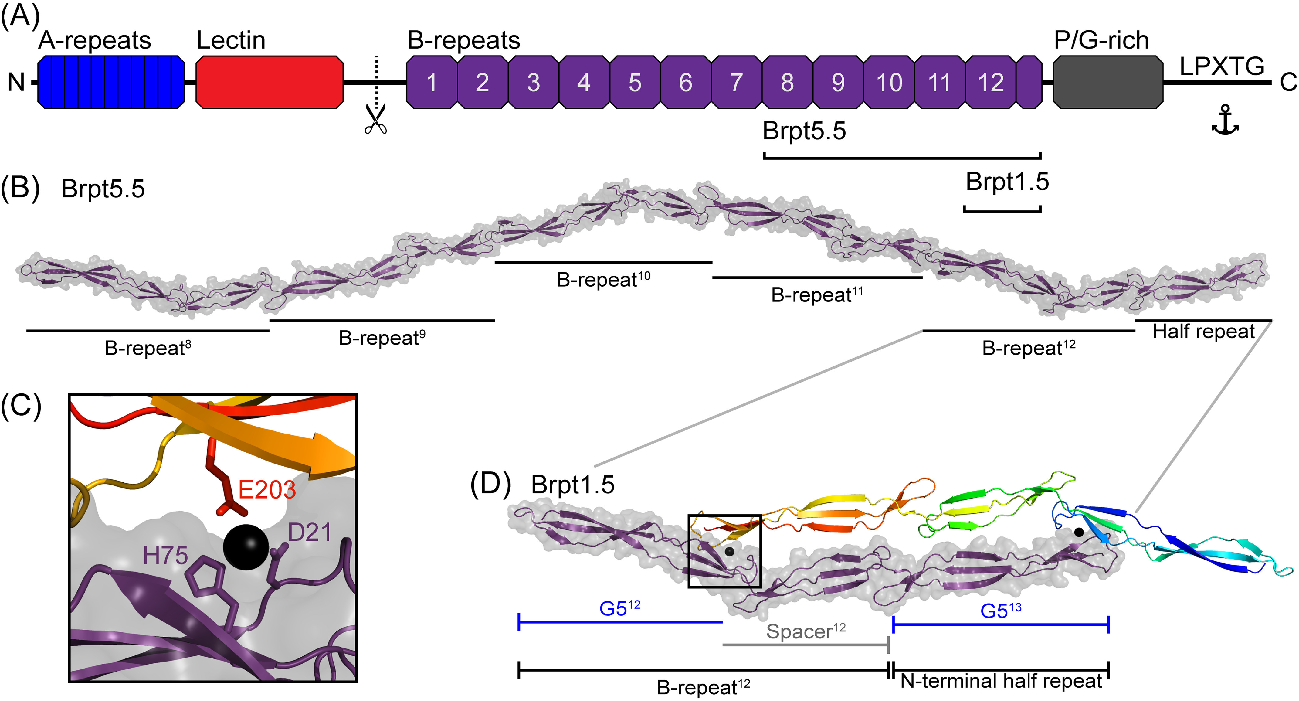
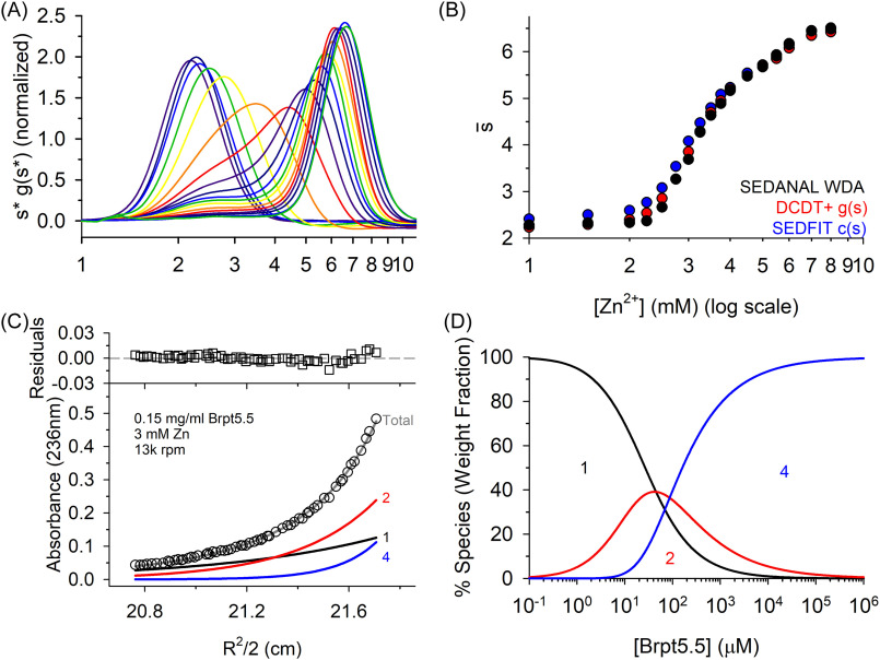
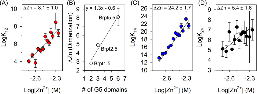
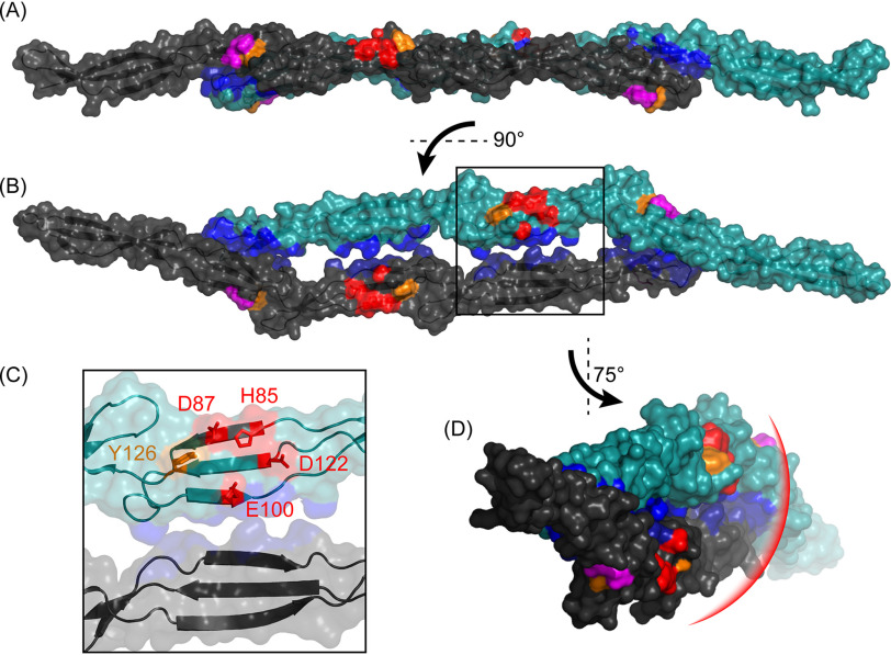
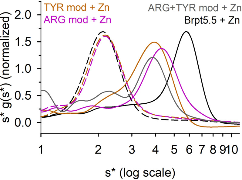
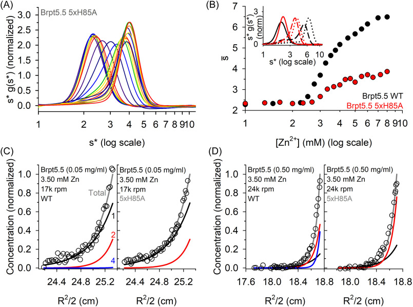
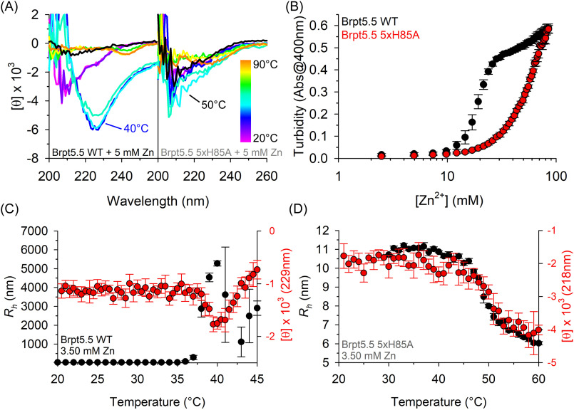
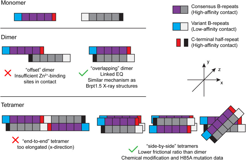
The accumulation-associated protein (Aap) from Staphylococcus epidermidis is a biofilm-related protein that was found to be a critical factor for infection using a rat catheter model. The B-repeat superdomain of Aap, composed of 5-17 B-repeats, each containing a Zn2+-binding G5 and a spacer subdomain, is responsible for Zn2+-dependent assembly leading to accumulation of bacteria during biofilm formation. We previously demonstrated that a minimal B-repeat construct (Brpt1.5) forms an antiparallel dimer in the presence of 2-3 Zn2+ ions. More recently, we have reported the presence of functional amyloid-like fibrils composed of Aap within S. epidermidis biofilms and demonstrated that a biologically relevant construct containing five and a half B-repeats (Brpt5.5) forms amyloid-like fibrils similar to those observed in the biofilm. In this study, we analyze the initial assembly events of the Brpt5.5 construct. Analytical ultracentrifugation was utilized to determine hydrodynamic parameters of reversibly associating species and to perform linked equilibrium studies. Linkage studies indicated a mechanism of Zn2+-induced dimerization similar to smaller constructs; however, Brpt5.5 dimers could then undergo further Zn2+-induced assembly into a previously uncharacterized tetramer. This led us to search for potential Zn2+-binding sites outside of the dimer interface. We developed a Brpt5.5 mutant that was unable to form the tetramer and was concordantly incapable of amyloidogenesis. CD and dynamic light scattering indicate that a conformational transition in the tetramer species is a critical step preceding amyloidogenesis. This mechanistic model for B-repeat assembly and amyloidogenesis provides new avenues for potential therapeutic targeting of staphylococcal biofilms.

Keywords: analytical ultracentrifugation; biofilm; biophysics; chemical modification; oligomerization; protein aggregation; protein chemical modification; sedimentation equilibrium; thermodynamics.


Conflict of interest statement

Conflict of interest—A. B. H. serves as a Scientific Advisory Board member for Hoth Therapeutics, Inc., holds equity in Hoth Therapeutics and Chelexa BioSciences, LLC, and was a co-inventor on three patents broadly related to the subject matter of this work.

Figures

Figure 1.
Figure 1.
A, domain arrangement of Aap from S. epidermidis RP62A. The N terminus contains short repeats followed by a lectin domain. A proteolytic cleavage site allows for removal of this region during biofilm accumulation, exposing the B-repeat superdomain, a repetitive proline/glycine-rich region, and a cell wall–anchoring motif. B, a model of Brpt5.5 with each B-repeat labeled, with numbers corresponding to position in the scheme from A. C and D, structure of the Brpt1.5 dimer (with two Zn2+ ions shown as black spheres; Protein Data Bank entry 4FUN). The model of Brpt5.5 shown in B is constructed from the superposition of multiple Brpt1.5 models, with the N-terminal half-repeat overlaying the G5 domain from each of the other B-repeats. These images were generated using PyMOL (PyMOL Molecular Graphics System, version 1.8, Schrödinger, LLC).
Figure 2.
Figure 2.
Brpt5.5 exhibits a monomer-dimer-tetramer equilibrium in the presence of Zn2+. A, WDA of 0.50 mg/ml Brpt5.5 in the presence of increasing ZnCl2 concentrations, starting at 0 mm ZnCl2 at the left in purple, shifting to the right up to 8 mm ZnCl2 in green. B, the weight-average sedimentation coefficient (s¯) at increasing ZnCl2 concentrations. Data plotted were analyzed by separate programs, as designated in the key. The data presented in A and B are from a series of experiments using one batch of Brpt5.5 WT. At least three replicates were performed at 3.50, 5.00, and 8 mm ZnCl2, providing results within 0.5 S of the presented data. C, a representative sedimentation equilibrium AUC data set at 3 mm Zn2+, 0.15 mg/ml Brpt5.5, at 13,000 rpm. This data set is part of a global fit of six or more curves (at least three protein concentrations and at least three speeds). Empty circles, raw absorbance data at 236 nm; solid gray line, best fit, with residuals shown in the top plot. Individual species are represented by lines in black (monomer), red (dimer), and blue (tetramer). D, distribution of each species based on Brpt5.5 concentration (6.5 μm = 0.50 mg/ml), calculated from the determined association constants at 3 mm Zn2+. The x axis extends until saturation of monomer or tetramer.
Figure 3.
Figure 3.
Analysis of linked equilibria reveals the number of Zn2+ ions bound during each assembly event. A, Wyman plot of linked equilibria for the dimer. The slope of the weighted linear regression indicates the number of Zn2+ ions bound during dimerization (ΔZn = 8.1 ± 1.0). B, a comparison of the ΔZn values per G5 domain for Brpt5.5 (determined in this study) with Brpt2.5 and Brpt1.5 (previously published by Conrady et al. (9)). The Wyman plot for the tetramer is shown based on the overall monomer-tetramer association constant (K14) in C and the stepwise dimer-tetramer association constant (K24) in D. Error bars for A, C, and D represent the 95% confidence intervals for fitted association constants. The error bar for Brpt5.5 data in B represents the range of ΔZn for Brpt5.5 dimerization reported in A.
Figure 4.
Figure 4.
Chemical modification targets and potential Zn2+-binding residues are highlighted on a structure of Brpt1.5 dimer (Protein Data Bank entry 4FUN). Tyrosine residues are colored orange, arginine residues are colored magenta, and hypothesized Zn2+-binding residues important in tetramer formation are colored red. The dimer interface surface is colored blue. A is rotated 90° along the y axis compared with B, which is turned 75° to create a view looking down along the side of the dimer in D. The bottom left inset (C) shows greater detail in the region within the black square from B. The red arc in the bottom right image (D) represents where we would expect a second dimer to interface with the presented dimer. These images were generated using PyMOL (The PyMOL Molecular Graphics System, version 1.8, Schrödinger, LLC).
Figure 5.
Figure 5.
Chemical modifications inhibit tetramer formation. Sedimentation velocity AUC data were analyzed by WDA. Chemical modification of Tyr (orange), Arg (magenta), and both types of residues (gray) reduces assembly in the presence of 5 mm ZnCl2 (solid lines) compared with unmodified Brpt5.5 (black). Modification of Brpt5.5 caused no change in sedimentation of the monomer in the absence of ZnCl2 (dashed lines).
Figure 6.
Figure 6.
H85A mutations inhibit tetramer formation. A, sedimentation velocity AUC data analyzed by WDA for the Brpt5.5 5xH85A mutant. B compares the weight-average sedimentation coefficient of WT and 5xH85A at various ZnCl2 concentrations as determined by WDA. 5xH85A displays only one sigmoidal transition plateauing at ∼4 S. An inset in B shows distributions of WT (black) and 5xH85A (red) without ZnCl2 (solid lines), with 3.50 mm ZnCl2 (dashed lines), or with 5.00 mm ZnCl2 (dotted lines). The data in A and B originate from one complete data set. Single experiments have also been performed at 3.50, 5.00, and 8.00 mm ZnCl2 at least twice and provided results within 0.5 S of the presented data. C, sedimentation equilibrium AUC data for Brpt5.5 WT and 5xH85A at the lowest protein-loading concentration (0.05 mg/ml). At this loading concentration, WT produces very little tetramer (blue line) but significant amounts of dimer (red line). Comparing the left (WT) and right (5xH85A) halves of this panel shows how similar the dimerization behavior is between the two proteins. D, however, is at 0.50 mg/ml Brpt5.5, a condition producing significant tetramer in WT (left). Under the same conditions, 5xH85A (right) showed only monomer and dimer. Empty black circles, raw absorbance data; gray lines, fits for total signal; black, red, and blue lines, monomer, dimer, and tetramer species fits.
Figure 7.
Figure 7.
Inhibiting tetramer formation results in weaker aggregation propensity. A, far-UV wavelength scans at 10 °C increments, from 20 to 90 °C. The black spectra represent scans taken at 20 °C, after the sample had been heated to 90 °C. Brpt5.5 WT shows significant change in the Brpt5.5 CD signal at 40 °C in the presence of Zn2+ (specifically ∼225 nm), whereas the 5xH85A mutant does not show this behavior. B examines the turbidity of Brpt5.5 WT (black circles) or 5xH85A (red circles) upon Zn2+ additions. Data points plotted are averages from three replicate experiments, with error bars showing S.D. C and D, the Rh measured by DLS (black circles) overlaid with CD data collected at a single wavelength (red circles; wavelength specified on the right y axis). DLS data points are averages from triplicate measurements at each temperature during a single experiment. DLS error bars show ± 1 S.D., whereas CD error bars are the error reported by the instrument. The DLS data sets presented are representative of three experiments performed at 0.5–1 mg/ml in 3.50 mm ZnCl2, whereas CD data sets shown are representative of at least two experiments performed at 0.5–1 mg/ml with 3.50 mm ZnCl2 or 5 mm ZnCl2.
Figure 8.
Figure 8.
Models of tandem B-repeat reversible assembly. Each B-repeat can be described as “Consensus” (purple and gray) or “Variant” (blue and white) according to Shelton et al. (13). Brpt5.5 contains one variant and four consensus B-repeats, in addition to the C-terminal half-repeat cap. Consensus B-repeats form high-affinity contacts with one another in the presence of Zn2+, whereas variant B-repeats form low-affinity contacts. The C-terminal half-repeat is a G5 domain that behaves similarly to the G5 domain in consensus B-repeats. Based on the results of this study, we can eliminate several models of assembly based on biophysical data. However, we will require additional data to distinguish between the side-by-side tetramer configurations.

Similar articles

Cited by

References

    1. Otto M. (2009) Staphylococcus epidermidis—the “accidental” pathogen. Nat. Rev. Microbiol. 7, 555–567 10.1038/nrmicro2182 - DOI - PMC - PubMed
    1. Otto M. (2008) Staphylococcal biofilms. Curr. Top. Microbiol. Immunol. 322, 207–228 10.1007/978-3-540-75418-3_10 - DOI - PMC - PubMed
    1. National Nosocomial Infections Surveillance System (2004) National Nosocomial Infections Surveillance (NNIS) System Report, data summary from January 1992 through June 2004, issued October 2004. Am. J. Infect. Control 32, 470–485 10.1016/j.ajic.2004.10.001 - DOI - PubMed
    1. Donlan R. M. (2001) Biofilms and device-associated infections. Emerg. Infect. Dis. 7, 277–281 10.3201/eid0702.010226 - DOI - PMC - PubMed
    1. Costerton J. W., Stewart P. S., and Greenberg E. P. (1999) Bacterial biofilms: a common cause of persistent infections. Science 284, 1318–1322 10.1126/science.284.5418.1318 - DOI - PubMed

Publication types

MeSH terms

Associated data

LinkOut - more resources