. 2020 Jul 28;117(30):17510-17512.
doi: 10.1073/pnas.1921324117. Epub 2020 Jul 14.

Type I IFN is siloed in endosomes

Affiliations

Type I IFN is siloed in endosomes

Jennie B Altman et al. Proc Natl Acad Sci U S A. .

Abstract

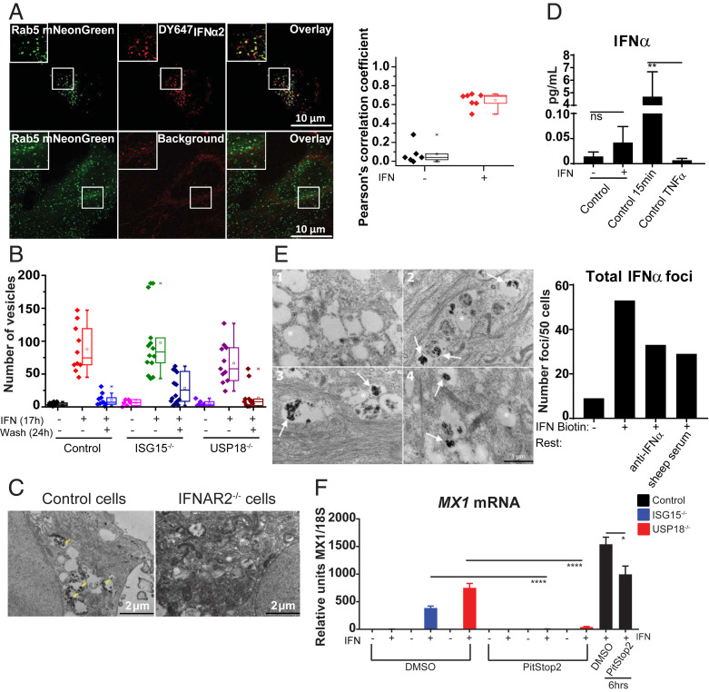
Type I IFN (IFN-I) is thought to be rapidly internalized and degraded following binding to its receptor and initiation of signaling. However, many studies report the persistent effects mediated by IFN-I for days or even weeks, both ex vivo and in vivo. These long-lasting effects are attributed to downstream signaling molecules or induced effectors having a long half-life, particularly in specific cell types. Here, we describe a mechanism explaining the long-term effects of IFN-I. Following receptor binding, IFN-I is siloed into endosomal compartments. These intracellular "IFN silos" persist for days and can be visualized by fluorescence and electron microscopy. However, they are largely dormant functionally, due to IFN-I-induced negative regulators. By contrast, in individuals lacking these negative regulators, such as ISG15 or USP18, this siloed IFN-I can continue to signal from within the endosome. This mechanism may underlie the long-term effects of IFN-I therapy and may contribute to the pathophysiology of type I interferonopathies.

Keywords: cytokine retention; endosome; type I interferon.


Conflict of interest statement

The authors declare no competing interest.

Figures

Fig. 1.
Fig. 1.
Persistence of IFN-I signaling in ISG15- and USP18-deficient cells. (A) Control, ISG15-, or USP18-deficient fibroblasts were given 0.197 nM (1,000 u/mL) IFNα2b for 12 h, washed, and rested for 36 h in all experiments. Relative units of MX1 mRNA was quantified by qPCR. (B) Following A, EU was added for 24 h, RNA containing EU isolated (nascent RNA), and qPCR performed. (C and D) Western blotting or IF for phospho-STAT1, phospho-STAT2, and total STAT2 as per A. Band densitometry of pSTAT1 relative to tubulin for C. (Magnification in D: 40×.) (E) Cells were incubated with 10 μM Cerdulatinib (Cerd) or vehicle (dimethyl sulfoxide [DMSO]) for 4 h following A, and Western blotting was performed. (F and G) Cells were primed and rested with antibodies present, and (F) qPCR or (G) Western blotting was performed. (H) qPCR for IFNB mRNA after prime-rest protocol or polyinosinic:polycytidylic acid (poly I:C). (I) Cells were prime-rested as per A, and supernatants were placed on naïve control cells for 8 h (NT) or with anti-IFNα. Control cells given 0.197 nM IFN-I (8 h) served as a positive control, and RSAD2 mRNA was quantified. SEM represented, unpaired Student t tests: *P < 0.05, **P < 0.01, ***P < 0.001, ****P < 0.0001. ns = not significant.
Fig. 2.
Fig. 2.
IFN-I is retained intracellularly. (A) HeLa cells expressing Rab5-mNeonGreen (green) incubated with 5 nM DY647IFNα2 (magenta) for 17 h, washed, rested for 25 h, and visualized by LLSM (Top). Untreated cells show background autofluorescence (Bottom). Z projections are of five sections (0.52-µm total thickness). Box plot shows correlation coefficients for colocalization of IFNα2 and Rab5 (n = 10 cells). (B) Control, ISG15-, and USP18-deficient cells were given DY647IFNα2 for 17 h and imaged, or rested for 24 h and IFN-I vesicles quantified. (C) EM of Control or IFNAR2-deficient cells given 0.5 nM of IFN-I biotin for 15 min. Yellow arrows denote positive immunoreactivity. (D) Prime-rested cells evaluated by SiMoA, P = 0.0093. (E) EM of control cells given 0.5 nM of IFN-I biotin for 12 h, washed and rested in media containing 0.2 μg/mL anti-IFNa or vehicle (sheep serum) for 36 h. Representative example of (1) negative control without IFN-I biotin; (2) IFN-I biotin 12 h, 36 h rest; (3) IFN-I biotin 12 h, anti-IFNα during 36 h rest; and (4) IFN-I biotin 12 h, sheep serum during 36 h rest. Asterisk denotes unlabeled endosome; arrow denotes positive immunoreactivity within endosome; n = 50 cells per condition. (F) Cells were primed-rested in DMSO or 100 μM PitStop2, and qPCR was performed. Cells given IFN-I with DMSO or PitStop2 for 6 h served as controls. SEM represented, unpaired Student t tests: *P < 0.05, ****P < 0.0001.

References

    1. Borden E. C., et al. , Interferons at age 50: Past, current and future impact on biomedicine. Nat. Rev. Drug Discov. 6, 975–990 (2007). - PMC - PubMed
    1. Shechter Y., Preciado-Patt L., Schreiber G., Fridkin M., Prolonging the half-life of human interferon-alpha 2 in circulation: Design, preparation, and analysis of (2-sulfo-9-fluorenylmethoxycarbonyl)7- interferon-alpha 2. Proc. Natl. Acad. Sci. U.S.A. 98, 1212–1217 (2001). - PMC - PubMed
    1. Brennan B. J., Xu Z. X., Grippo J. F., Evaluation of the absolute bioavailability of pegylated interferon alfa-2a after subcutaneous administration to healthy male volunteers: An open-label, randomized, parallel-group study. Clin. Ther. 34, 1883–1891 (2012). - PubMed
    1. Lai L., Hui C. K., Leung N., Lau G. K., Pegylated interferon alpha-2a (40 kDa) in the treatment of chronic hepatitis B. Int. J. Nanomedicine 1, 255–262 (2006). - PMC - PubMed
    1. Hermesh T., Moltedo B., Moran T. M., López C. B., Antiviral instruction of bone marrow leukocytes during respiratory viral infections. Cell Host Microbe 7, 343–353 (2010). - PMC - PubMed

Publication types

LinkOut - more resources