. 2020 Jul 14;10(1):11540.
doi: 10.1038/s41598-020-68405-4.

Uranium-free X solution: a new generation contrast agent for biological samples ultrastructure

Affiliations

Uranium-free X solution: a new generation contrast agent for biological samples ultrastructure

Aldo Moscardini et al. Sci Rep. .

Abstract

Biological samples are mainly composed of elements with a low atomic number which show a relatively low electron scattering power. For Transmission Electron Microscopy analysis, biological samples are generally embedded in resins, which allow thin sectioning of the specimen. Embedding resins are also composed by light atoms, thus the contrast difference between the biological sample and the surrounding resin is minimal. Due to that reason in the last decades, several staining solutions and approaches, performed with heavy metal salts, have been developed with the purpose of enhancing both the intrinsic sample contrast and the differences between the sample and resin. The best staining was achieved with the uranyl acetate (UA) solution, which has been the election method for the study of morphology in biological samples. More recently several alternatives for UA have been proposed to get rid of its radiogenic issues, but to date none of these solutions has achieved efficiencies comparable to UA. In this work, we propose a different staining solution (X Solution or X SOL), characterized by lanthanide polyoxometalates (LnPOMs) as heavy atoms source, which could be used alternatively to UA in negative staining (NS), in en bloc staining, and post sectioning staining (PSS) of biological samples. Furthermore, we show an extensive chemical characterization of the LnPOM species present in the solution and the detailed work for its final formulation, which brought remarkable results, and even better performances than UA.


Conflict of interest statement

The authors declare no competing interests.

Figures
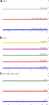
Figure 1
Figure 1
NMR study of complex formation and stability: (a) 31P-NMR titration of PTA with increasing amounts of YbCl3. (b) 31P-NMR spectra of the active specie in a range of YbCl3/PTA molar ratios. (c) 31P-NMR spectra of a 1:1 mixture of PTA and YbCl3 in a pH = 2–7 range.
Figure 2
Figure 2
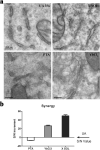
PTA and YbCl3 synergy: (a) EM micrographs of Mia PaCa-2 cells stained with UA 3% solution, X SOL, PTA 3.2 mM, YbCl3 48 mM (all solutions are made in water/ethanol 80/20 v/v); (b) quantitative analysis of the staining efficiency on Mia PaCa-2 cells.
Figure 3
Figure 3
Buffering: (a) EM micrographs of gastrocnemius muscle stained with UA 3% solution or X SOL (upper line). The staining seems much more uniform in UA 3% stained samples compared to X SOL stained one; higher magnification of X SOL stained muscle (boxed lower line) shows that differences in contrast is due to the contraction status of myofibers, thus discriminating between relaxed and contracted ones; (b) Quantitative analysis of the staining efficiency performed on Mia PaCa-2 cells; (c) 31P-NMR analysis of X SOL w/wo the addition of MES buffer.
Figure 4
Figure 4
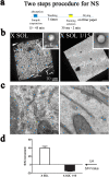
NS and dilution: (a) representative scheme of the two steps protocol for NS; (b) EM micrographs of synthetic liposomes negatively stained with X Solution, pure or diluted in water 1/15 (V/V); (c) EM micrographs of Mia PaCa-2 cells en bloc stained with X Solution, pure or diluted in water 1/15 (V/V); (d) Quantitative analysis of the staining efficiency performed on Mia PaCa-2 cells.
Figure 5
Figure 5
Mechanistic hypothesis of X Solution staining process: (a) Clathrin coated vesicles formation in embedded Mia PaCa-2 cells en bloc stained with X SOL; X Solution clearly shows clathrin shells in the cytosolic side, cargo protein in the extracellular side, and, also, proteins’ transmembrane domains (white arrows); (b) 31P-NMR of the interaction between a mixed solution of mono-and dibuthylphosphate: alone or associated with PTA, YbCl3 and X Solution.

References

    1. Watson ML. Staining of tissue sections for electron microscopy with heavy metals. J. Biophys. Biochem. Cytol. 1958;4:475–478. doi: 10.1083/jcb.4.4.475. - DOI - PMC - PubMed
    1. Terzakis JA. Uranyl acetate, a stain and a fixative. J. Ultrastruct. Res. 1968;22:168–184. doi: 10.1016/S0022-5320(68)90055-5. - DOI - PubMed
    1. Nakakoshi M, Nishioka H, Katayama E. New versatile staining reagents for biological transmission electron microscopy that substitute for uranyl acetate. J. Electron Microsc. (Tokyo) 2011;60:401–407. doi: 10.1093/jmicro/dfr084. - DOI - PubMed
    1. Hosogi N, Nishioka H, Nakakoshi M. Evaluation of lanthanide salts as alternative stains to uranyl acetate. Microscopy. 2015;64:429–435. doi: 10.1093/jmicro/dfv054. - DOI - PubMed
    1. Benmeradi N, Payre B, Goodman SL. Easier and safer biological staining: High contrast UranyLess staining of TEM Grids using mPrep/g capsules. Microsc. Microanal. 2015;21:721–722. doi: 10.1017/S1431927615004407. - DOI