. 2020 Jul 14;10(1):11604.
doi: 10.1038/s41598-020-68308-4.

Region-specific mechanisms of corticosteroid-mediated inotropy in rat cardiomyocytes

Affiliations

Region-specific mechanisms of corticosteroid-mediated inotropy in rat cardiomyocytes

Caroline Wacker et al. Sci Rep. .

Abstract

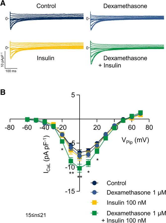
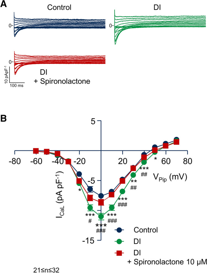
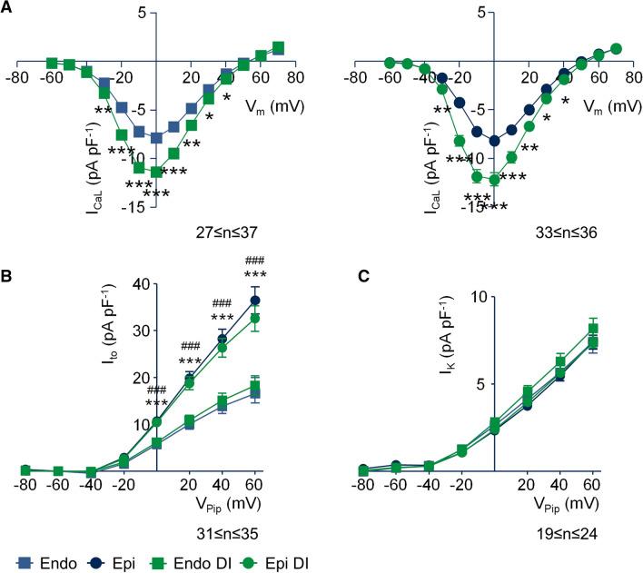
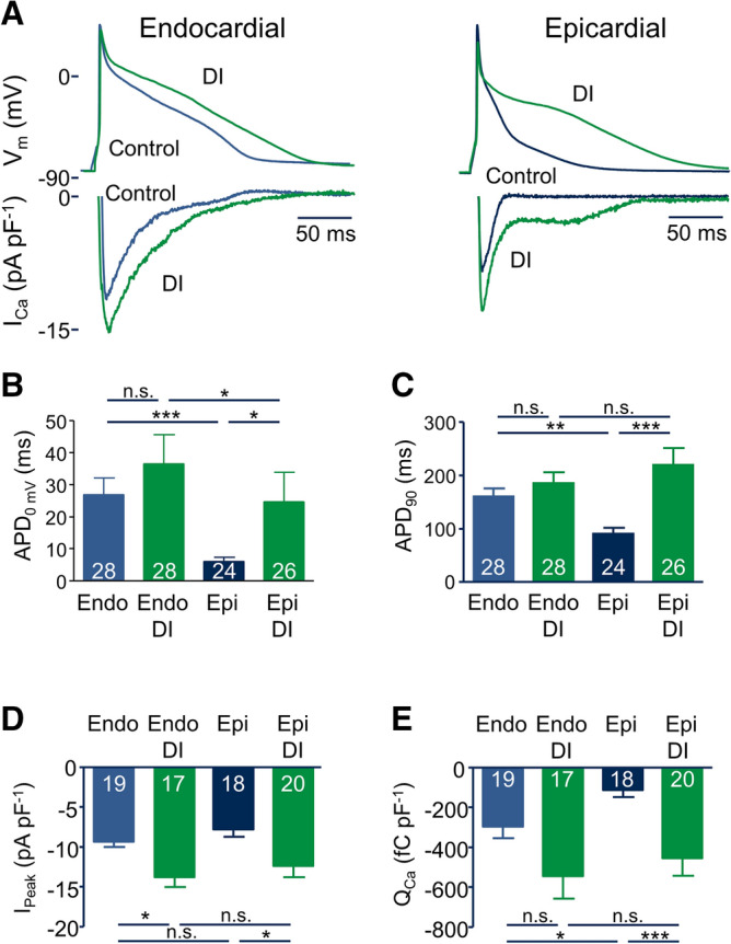
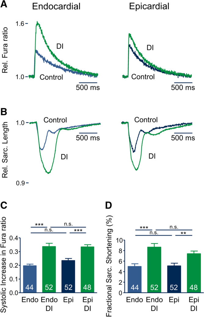
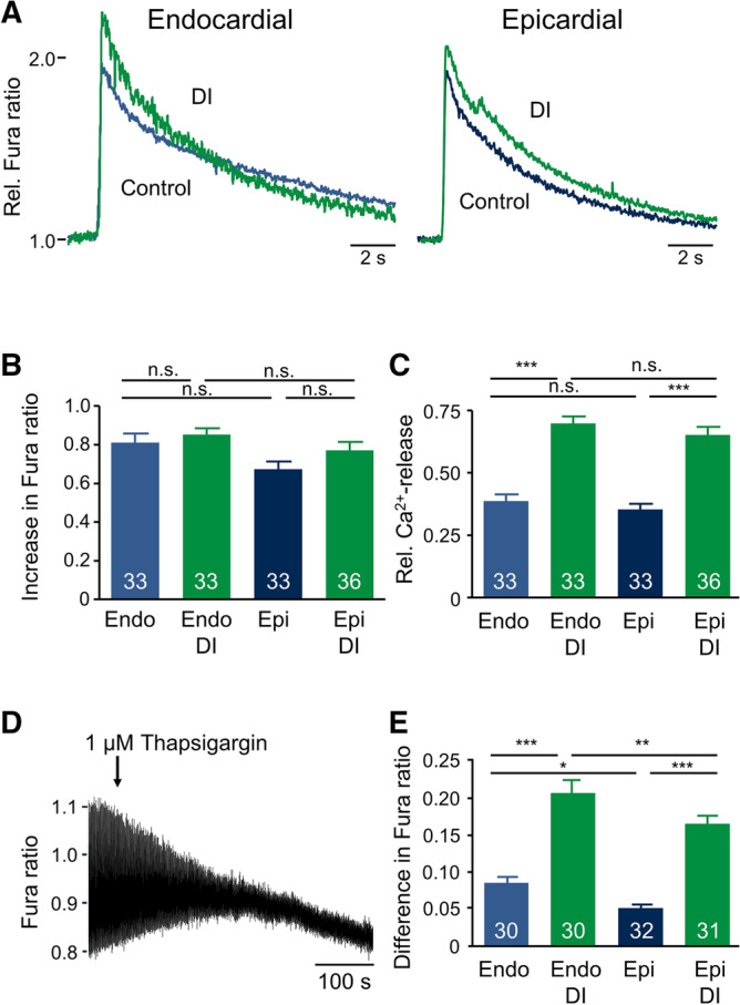
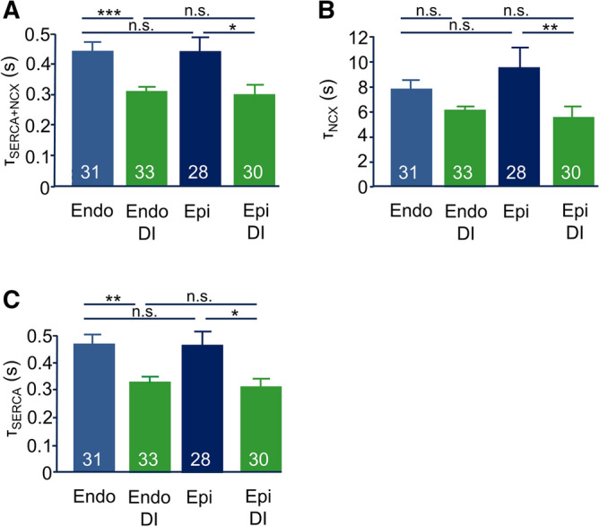
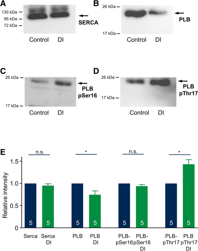
Regional differences in ion channel activity in the heart control the sequence of repolarization and may contribute to differences in contraction. Corticosteroids such as aldosterone or corticosterone increase the L-type Ca2+ current (ICaL) in the heart via the mineralocorticoid receptor (MR). Here, we investigate the differential impact of corticosteroid-mediated increase in ICaL on action potentials (AP), ion currents, intracellular Ca2+ handling and contractility in endo- and epicardial myocytes of the rat left ventricle. Dexamethasone led to a similar increase in ICaL in endocardial and epicardial myocytes, while the K+ currents Ito and IK were unaffected. However, AP duration (APD) and AP-induced Ca2+ influx (QCa) significantly increased exclusively in epicardial myocytes, thus abrogating the normal differences between the groups. Dexamethasone increased Ca2+ transients, contractility and SERCA activity in both regions, the latter possibly due to a decrease in total phospholamban (PLB) and an increase PLBpThr17. These results suggest that corticosteroids are powerful modulators of ICaL, Ca2+ transients and contractility in both endo- and epicardial myocytes, while APD and QCa are increased in epicardial myocytes only. This indicates that increased ICaL and SERCA activity rather than QCa are the primary drivers of contractility by adrenocorticoids.


Conflict of interest statement

The authors declare no competing interests.

Figures

Figure 1
Figure 1
Dexamethasone increases ICaL only in the presence of insulin. (A) Representative whole-cell current traces of ICaL recorded from myocytes incubated for 24 h under control conditions (Control), with 1 µM dexamethasone, 100 nM insulin, 1 µM dexamethasone + 100 nM insulin. Myocytes were clamped for 600 ms from the holding potential of VPip = − 90 mV to test potentials between VPip = − 60 mV up to + 70 mV in steps of 10 mV. Na+ currents were inactivated by a prepulse of 70 ms to − 50 mV. Basic cycle length was 3,000 ms. (B) Average current–voltage relations of currents similar to those shown in A. ICaL was quantified by subtracting the peak current from the current at the end of the voltage pulse (at 600 ms). *p < 0.05, **p < 0.01, dexamethasone + insulin versus control. 15 ≤ n ≤ 21.
Figure 2
Figure 2
The effect of dexamethasone is mediated by the mineralocorticoid receptor. (A) Representative whole-cell current traces of ICaL recorded from myocytes incubated for 24 h under control conditions (Control), with 1 µM dexamethasone + 100 nM insulin and 1 µM dexamethasone + 100 nM insulin + 10 µM spironolactone. Myocytes were clamped for 600 ms from the holding potential of VPip = − 90 mV to test potentials between VPip = − 60 mV up to + 70 mV in steps of 10 mV. Basic cycle length was 3,000 ms. (B) Average current–voltage relations of currents similar to those shown in (A). ICaL was quantified by subtracting the peak current from the current at the end of the voltage pulse (at 600 ms). *p < 0.05, **p < 0.01, ***p < 0.001, dexamethasone + insulin versus control; #p < 0.05, ##p < 0.01, ###p < 0.001 dexamethasone + insulin versus dexamethasone + insulin + spironolactone. 21 ≤ n ≤ 32.
Figure 3
Figure 3
Effect of DI treatment on ICaL and K+ currents in endo- and epicardial myocytes. (A) Average current–voltage relations of recordings similar to those shown in Fig. 1 and 2, obtained from endo- and epicardial myocytes of the left ventricular free wall incubated for 24 h under control conditions (blue) and with DI (green). *p < 0.05, **p < 0.01, ***p < 0.001, DI versus control. (B and C) Average current–voltage relations of Ito and IK recorded from endo- and epicardial myocytes of the left ventricular free wall incubated for 24 h under control conditions (blue) and DI (green). Myocytes were clamped for 600 ms from the holding potential of VPip = − 90 mV to test potentials between VPip =  + 60 mV to − 80 mV in steps of −20 mV. Basic cycle length was 3,000 ms. Ito was quantified by subtracting the peak current from the current at the end of the voltage pulse (at 600 ms), IK was estimated as the current at the end of the voltage pulse (600 ms). ***p < 0.001, epi- versus endocardial myocytes incubated under control conditions, ###p < 0.001, epi- versus endocardial myocytes incubated with DI.
Figure 4
Figure 4
DI treatment abrogates the gradient in APD. ( A) Representative APs and corresponding AP-induced Ca2+ currents recorded from endo- and epicardial myocytes incubated for 24 h under control conditions (blue) and with DI (green). APs were elicited by a train of suprathreshold depolarizing current injections at a basic cycle length of 1,000 ms. (B) Average APD at repolarization to 0 mV (APD0mV). (C) Average APD at 90% repolarization (APD90). (D) Average peak Ca2+ influx (IPeak) and (E) average total AP-induced Ca2+ influx (QCa) obtained from similar recordings to those shown in ( A). Numbers in bars indicate number of myocytes in each group.
Figure 5
Figure 5
Effect of DI treatment on Ca2+ transients and contractility in endo- and epicardial myocytes. (A) Representative Ca2+ transient after 24 h incubation under control conditions (blue curve) and with DI (green curve). (B) Representative sarcomere length recordings of the same myocytes shown in A. C, average systolic increase in Fura ratio of recordings similar to those shown in (A), (D) average fractional sarcomere shortening of recordings similar to those shown in A. Numbers in bars indicate number of myocytes in each group.
Figure 6
Figure 6
Effect of DI treatment on SR Ca2+ handling. (A) Representative caffeine-induced Ca2+ transients after 24 h incubation under control conditions (blue curves) and with DI (green curves). Caffeine (10 mM) was applied after 60 s of pacing at 1 Hz to ensure equal SR loading. (B) Average increase in Fura ratio in response to caffeine application. (C) Average relative Ca2+ release (Rel. Ca2+-release) calculated by dividing the increase in Fura ratio during regular pacing (1 Hz) by the increase in Fura ratio after application of caffeine. (D) Representative recording of Ca2+ transients after application of 1 µM thapsigargin obtained from a myocyte incubated under control conditions. (E) Average difference of systolic increase in Fura ratio calculated by subtraction of the systolic increase in Fura ratio after 5 min incubation with 1 µM thapsigargin from the systolic increase in Fura ratio before application of 1 µM thapsigargin. Numbers in bars indicate number of myocytes in each group.
Figure 7
Figure 7
Effect of DI treatment on the time constants of cytosolic Ca2+ removal pathways. (A) Average Ca2+ transport time constants of SERCA + NCX (τSERCA + NCX) estimated by monoexponential fitting of the decay of the Ca2+ transients recorded during regular pacing at 1 Hz. (B) Average Ca2+ transport time constants of NCX (τNCX) obtained by monoexponential fitting of the decay of the Ca2+ transients in response to caffeine application. (C) Average average Ca2+ transport time constants of SERCA (τSERCA) calculated by subtracting the NCX-rate constant calculated from caffeine-induced Ca2+ transients from the rate constants of the decay of Ca2+ transient during regular pacing. Numbers in bars indicate number of myocytes in each group.
Figure 8
Figure 8
Effect of DI treatment on SERCA and PLB protein expression and phosphorylation. (A-D) Representative western blots stained for SERCA2 (A, 100 kDa), PLB (B, 25 kDa), phospho-PLB-Ser16 (C, 25 kDa) and phospho-PLB-Thr17 (D, 25 kDa). Protein was isolated from left ventricular myocytes incubated for 24 h under control conditions (Control) or with DI. Arrows indicate the according molecular weight level of the target protein. (E) Average relative protein expression was assessed by western blot quantification (normalization by Ponceau staining) of control (blue) and DI treated myocytes (green). n = 5 matched cell isolations. The full length gels of the western blots including the corresponding Ponceau stains are shown in the supplemental figure.

References

    1. Bandulik S. Of channels and pumps: different ways to boost the aldosterone? Acta Physiol. (Oxf.) 2017;220:332–360. - PubMed
    1. Pitt B, et al. The effect of spironolactone on morbidity and mortality in patients with severe heart failure. Randomized Aldactone Evaluation Study Investigators. N. Engl. J. Med. 1999;341:709–717. - PubMed
    1. Pitt B, et al. Eplerenone, a selective aldosterone blocker, in patients with left ventricular dysfunction after myocardial infarction. N. Engl. J. Med. 2003;348:1309–1321. - PubMed
    1. Rubart M, Zipes DP. Mechanisms of sudden cardiac death. J. Clin. Invest. 2005;115:2305–2315. - PMC - PubMed
    1. Boixel C, Gavillet B, Rougier JS, Abriel H. Aldosterone increases voltage-gated sodium current in ventricular myocytes. Am. J. Physiol. Heart Circ. Physiol. 2006;290:H2257–H2266. - PubMed

Publication types

MeSH terms