. 2020 Jul 14;10(1):11566.
doi: 10.1038/s41598-020-68501-5.

High resolution MRI for quantitative assessment of inferior alveolar nerve impairment in course of mandible fractures: an imaging feasibility study

Affiliations

High resolution MRI for quantitative assessment of inferior alveolar nerve impairment in course of mandible fractures: an imaging feasibility study

Egon Burian et al. Sci Rep. .

Abstract

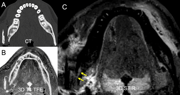
The purpose of this study was to evaluate a magnetic resonance imaging (MRI) protocol for direct visualization of the inferior alveolar nerve in the setting of mandibular fractures. Fifteen patients suffering from unilateral mandible fractures involving the inferior alveolar nerve (15 affected IAN and 15 unaffected IAN from contralateral side) were examined on a 3 T scanner (Elition, Philips Healthcare, Best, the Netherlands) and compared with 15 healthy volunteers (30 IAN in total). The sequence protocol consisted of a 3D STIR, 3D DESS and 3D T1 FFE sequence. Apparent nerve-muscle contrast-to-noise ratio (aNMCNR), apparent signal-to-noise ratio (aSNR), nerve diameter and fracture dislocation were evaluated by two radiologists and correlated with nerve impairment. Furthermore, dislocation as depicted by MRI was compared to computed tomography (CT) images. Patients with clinically evident nerve impairment showed a significant increase of aNMCNR, aSNR and nerve diameter compared to healthy controls and to the contralateral side (p < 0.05). Furthermore, the T1 FFE sequence allowed dislocation depiction comparable to CT. This prospective study provides a rapid imaging protocol using the 3D STIR and 3D T1 FFE sequence that can directly assess both mandible fractures and IAN damage. In patients with hypoesthesia following mandibular fractures, increased aNMCNR, aSNR and nerve diameter on MRI imaging may help identify patients with a risk of prolonged or permanent hypoesthesia at an early time.


Conflict of interest statement

The authors declare no competing interests.

Figures

Figure 1
Figure 1
This figure shows the acquired measurements for fracture dislocation in CT and 3D T1 FFE “bone sequence” (A). Nerve diameters showed a significant increase proximal of the fracture location (B). Further, it could be revealed that aNMCNR (C) and aSNR (D) were significantly higher at the fractured site compared to the healthy controls and to the contralateral side. (B) *p = 0.004, **p = 0.005, (C) *p = 0.007, **p = 0.021, (D) *p = 0.040.
Figure 2
Figure 2
In this clinical case a slightly displaced mandible fracture is illustrated (red arrows). The CT (A) and the 3D T1 FFE sequence (C) revealed the osseous continuity disruption with involvement of the mandibular canal with comparable accuracy. However, no conclusions regarding the condition of the IAN can be drawn as it is not visualized sufficiently. The DESS sequence allows for precise depiction of the IAN (B). The STIR sequence shows an increase in signal intensity within the IAN as well as an enlarged nerve diameter (D). The nerve continuity is preserved (yellow arrows).
Figure 3
Figure 3
In this clinical case a highly displaced mandible fracture is illustrated. The CT (A) and the 3D T1 FFE sequence (B) revealed the osseous continuity disruption with involvement of the mandible canal with comparable accuracy. However, no conclusions regarding the condition of the IAN can be drawn as it is not visualized sufficiently. The STIR sequence shows an increase in signal intensity within the IAN as well as an enlarged nerve diameter (C). The nerve continuity is preserved (yellow arrows).

References

    1. Dreizin D, Nam AJ, Hirsch J, Bernstein MP. New and emerging patient-centered CT imaging and image-guided treatment paradigms for maxillofacial trauma. Emerg. Radiol. 2018;25:533–545. doi: 10.1007/s10140-018-1616-9. - DOI - PubMed
    1. Ellis E., 3rd Treatment methods for fractures of the mandibular angle. Int. J. Oral Maxillofac. Surg. 1999;28:243–252. doi: 10.1016/S0901-5027(99)80152-0. - DOI - PubMed
    1. Ellis E., 3rd A prospective study of 3 treatment methods for isolated fractures of the mandibular angle. J. Oral Maxillofac. Surg. 2010;68:2743–2754. doi: 10.1016/j.joms.2010.05.080. - DOI - PubMed
    1. Kim MY, Kim CH, Han SJ, Lee JH. A comparison of three treatment methods for fractures of the mandibular angle. Int. J. Oral Maxillofac. Surg. 2016;45:878–883. doi: 10.1016/j.ijom.2016.02.013. - DOI - PubMed
    1. Tay AB, et al. Inferior alveolar nerve injury in trauma-induced mandible fractures. J. Oral Maxillofac. Surg. 2015;73:1328–1340. doi: 10.1016/j.joms.2015.02.003. - DOI - PubMed