Case Reports
. 2020 Jul 14;11(1):3572.
doi: 10.1038/s41467-020-17436-6.

Transplacental transmission of SARS-CoV-2 infection

Affiliations
Case Reports

Transplacental transmission of SARS-CoV-2 infection

Alexandre J Vivanti et al. Nat Commun. .

Abstract

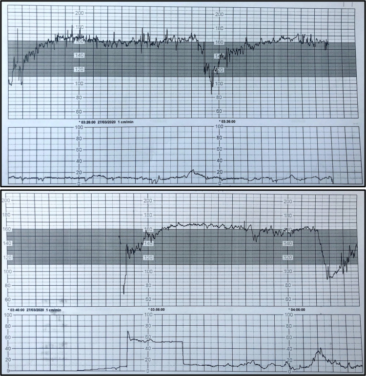
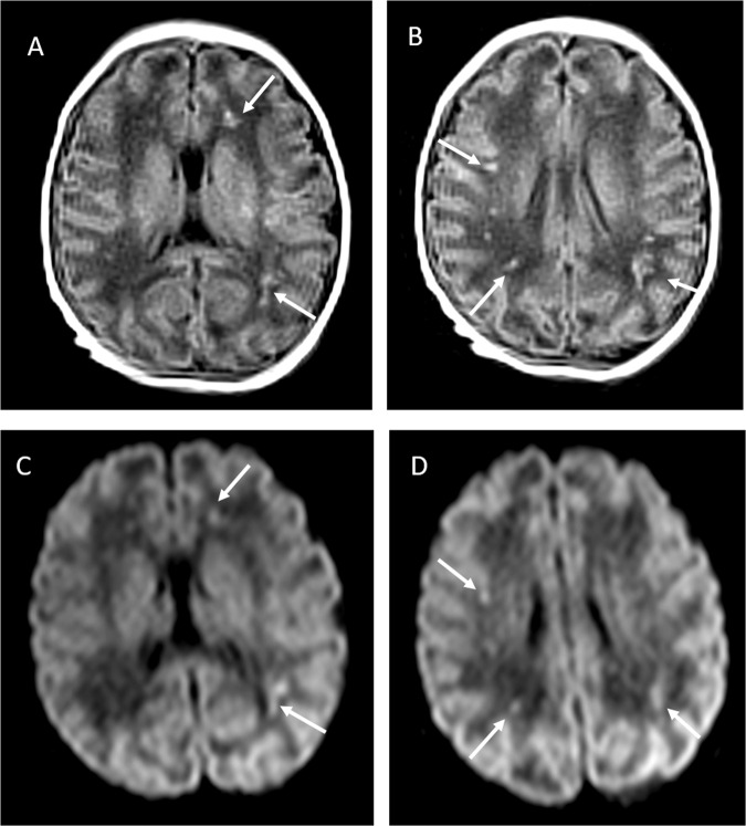
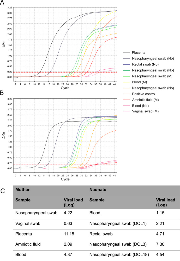
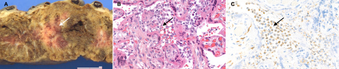
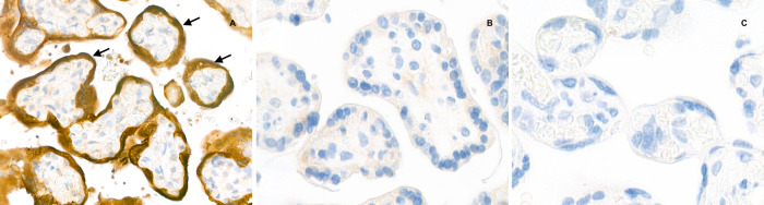
SARS-CoV-2 outbreak is the first pandemic of the century. SARS-CoV-2 infection is transmitted through droplets; other transmission routes are hypothesized but not confirmed. So far, it is unclear whether and how SARS-CoV-2 can be transmitted from the mother to the fetus. We demonstrate the transplacental transmission of SARS-CoV-2 in a neonate born to a mother infected in the last trimester and presenting with neurological compromise. The transmission is confirmed by comprehensive virological and pathological investigations. In detail, SARS-CoV-2 causes: (1) maternal viremia, (2) placental infection demonstrated by immunohistochemistry and very high viral load; placental inflammation, as shown by histological examination and immunohistochemistry, and (3) neonatal viremia following placental infection. The neonate is studied clinically, through imaging, and followed up. The neonate presented with neurological manifestations, similar to those described in adult patients.


Conflict of interest statement

The authors declare no competing interests.

Figures

Fig. 1
Fig. 1. Illustrative snapshot of fetal heart rate tracing.
Tachycardia, absent baseline variability, absence of accelerations with recurrent prolonged and late decelerations. These findings are highly suggestive of a pathological category III fetal heart rate tracing, which is strongly associated with adverse neonatal outcome. This cardiotogram was recorded 26 min before the cesarean section.
Fig. 2
Fig. 2. Cerebral MRI performed at 11 days of life.
a, b and c, d T1 and diffusion-weighted sequences, respectively. Images are taken at two different levels and show hyperintensities of the periventricular and subcortical frontal or parietal white matter (arrows).
Fig. 3
Fig. 3. Real-time polymerase chain reaction results.
a, b The E and S genes of SARS-CoV-2, respectively, for maternal and neonatal samples (X and Y axes represent the amount of amplified RNA and the number of cycles, respectively; the earlier the signal is detected, the lowest is the number of cycles and the higher the viral load is). c The viral load for each sample (expressed as Log copies/million of cells for the placenta and as Log copies/mL for all other specimens). All maternal samples were obtained right before the delivery or during C-section; newborn samples are listed chronologically and were obtained from the first to the third day of life, except for the last nasopharyngeal swab (obtained at 18 days of postnatal age). Colored lines represent the results of RT-PCR assay for each sample. The deep orange line represents the positive control, which is a SARS-CoV-2 culture supernatant (more details in “Methods”). Nasopharyngeal swabs at 1, 3 and 18 day of life are represented by the light orange, gray and green curves, respectively. Viral load in BAL fluidis not shown. DOL days of life, M maternal samples, Nb newborn samples.
Fig. 4
Fig. 4. Gross and microscopic examination of the placenta.
a The macroscopic lesions of perivillous fibrin deposition with infarction, as irregular strands of pale yellow-white induration (arrow). b Microscopic lesions of intervillositis characterized by an infiltrate of the intervillous spaces made of neutrophils and histiocytes (arrow) (HES stain, original magnification ×400). c The intervillositis with several CD68-positive histiocytes (arrow); neutrophils are negative with this anti-macrophage antibody (anti-CD68 immunohistochemistry, original magnification ×400).
Fig. 5
Fig. 5. Placental immunostaining for SARS-CoV-2 N-protein (anti-N immunohistochemistry, original magnification ×800).
a The intense brown cytoplasmic positivity of peri-villous trophoblastic cells in the placenta of our case (arrows). b, c Two negative controls (primary antibody, two SARS-CoV-2 negative placentas).

References

    1. Zeng H, et al. Antibodies in infants born to mothers with COVID-19 pneumonia. JAMA. 2020;323:1848–1849. - PMC - PubMed
    1. Dong L, et al. Possible vertical transmission of SARS-CoV-2 from an infected mother to her newborn. JAMA. 2020;323:1846–1848. - PMC - PubMed
    1. Zeng L, et al. Neonatal early-onset infection with SARS-CoV-2 in 33 neonates born to mothers with COVID-19 in Wuhan, China. JAMA Pediatr. 2020 doi: 10.1001/jamapediatrics.2020.0878. - DOI - PMC - PubMed
    1. Zhu H, et al. Clinical analysis of 10 neonates born to mothers with 2019-nCoV pneumonia. Transl. Pediatr. 2020;9:51–60. doi: 10.21037/tp.2020.02.06. - DOI - PMC - PubMed
    1. Yu N, et al. Clinical features and obstetric and neonatal outcomes of pregnant patients with COVID-19 in Wuhan, China: a retrospective, single-centre, descriptive study. Lancet Infect. Dis. 2020;20:559–564. doi: 10.1016/S1473-3099(20)30176-6. - DOI - PMC - PubMed

Publication types

MeSH terms