Review
. 2020 Sep;21(9):522-541.
doi: 10.1038/s41580-020-0262-8. Epub 2020 Jul 14.

The roles of histone variants in fine-tuning chromatin organization and function

Affiliations
Review

The roles of histone variants in fine-tuning chromatin organization and function

Sara Martire et al. Nat Rev Mol Cell Biol. 2020 Sep.

Abstract

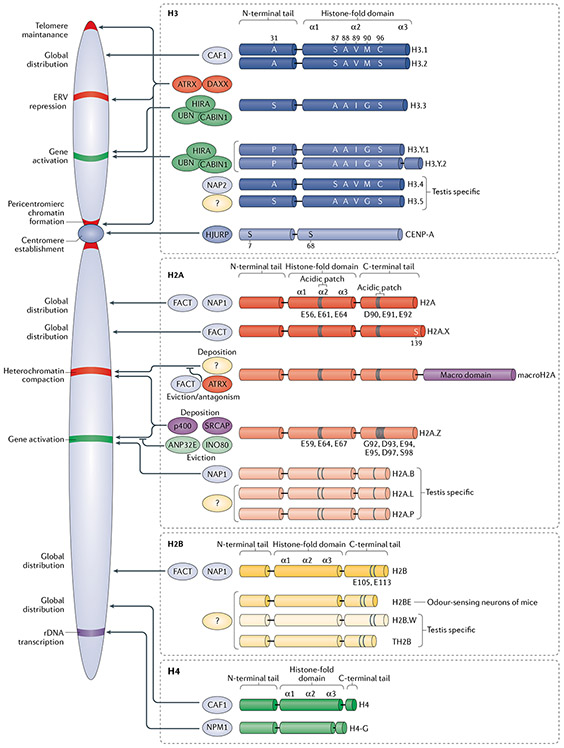
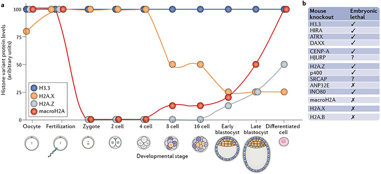
Histones serve to both package and organize DNA within the nucleus. In addition to histone post-translational modification and chromatin remodelling complexes, histone variants contribute to the complexity of epigenetic regulation of the genome. Histone variants are characterized by a distinct protein sequence and a selection of designated chaperone systems and chromatin remodelling complexes that regulate their localization in the genome. In addition, histone variants can be enriched with specific post-translational modifications, which in turn can provide a scaffold for recruitment of variant-specific interacting proteins to chromatin. Thus, through these properties, histone variants have the capacity to endow specific regions of chromatin with unique character and function in a regulated manner. In this Review, we provide an overview of recent advances in our understanding of the contribution of histone variants to chromatin function in mammalian systems. First, we discuss new molecular insights into chaperone-mediated histone variant deposition. Next, we discuss mechanisms by which histone variants influence chromatin properties such as nucleosome stability and the local chromatin environment both through histone variant sequence-specific effects and through their role in recruiting different chromatin-associated complexes. Finally, we focus on histone variant function in the context of both embryonic development and human disease, specifically developmental syndromes and cancer.


Figures

Fig. 1 ∣
Fig. 1 ∣. Core histones, their variants and associated chaperone/remodeller machineries.
Replication-coupled and variant core histones with respective chaperones, genomic distribution and functional output are shown. Specific amino acid residues are illustrated at either key differences among members of a common histone protein family (for example, histone H3.3 Ser31 vs H3.1 Ala31) or at well-established histone variant-specific post-translational modifications (for example, centromeric protein A (CENP-A) Ser7). Different shades of colour in the histone’s structures are used to indicate differences within the domains compared with the replication-coupled histone. See the text and TABLE 1 for additional details and references. ANP32E, acidic leucine-rich nuclear phosphoprotein 32 family member E; ATRX, α-thalassaemia mental retardation syndrome X-linked; CABIN1, calcineurin-binding protein cabin 1; CAF1, chromatin assembly factor 1; DAXX, death domain-associated protein; ERV, endogenous retroviral element; FACT, facilitates chromatin transcription; HIRA, histone regulator A; HJURP, Holliday junction recognition protein; NAP, nucleosome assembly protein; NPM1, nucleophosmin; rDNA, ribosomal DNA; UBN, ubinuclein; SRCAP, Snf2-related CREBBP CBP activator protein.
Fig. 2 ∣
Fig. 2 ∣. Variant-containing nucleosomes influence nucleosome stability and chromatin properties.
The sequence-specific histone variant properties together with selective incorporation of multiple histone variants into chromatin can influence (positively or negatively) nucleosome stability and result in different functional output on chromatin organization and function. Histone H2A.B lacks the acidic patch and leads to decreased nucleosome stability. By contrast, H2A.Z contains an extended acidic patch that contributes to increased nucleosome stability in homotypic H2A.Z nucleosomes. MacroH2A contains an extra domain and is also associated with increased nucleosome stability. Centromeric protein A (CENP-A) confers a much more rigid subnucleosomal structure through the presence of two extra amino acid residues (Arg80 (R80) and Gly81 (G81)) in the loop 1 (L1) region, which provide the connection between two major helices in H3 and CENP-A. Incorporation of histone H3.3 together with H2A.Z at promoters and enhancers results in the loosening of histone–histone and histone–DNA interactions, leading to chromatin opening, transcription factor (TF) recruitment and promotion of transcription. H2A.Z nucleosomes also contribute to chromatin compaction. In this case, H2A.Z nucleosomes together with heterochromatin-associated H3K9me3 promote chromatin recruitment and stabilization of heterochromatin protein 1α (HPα), which coordinates chromatin compaction and transcriptional gene silencing. Furthermore, H2A.Z and macroH2A nucleosomes are both involved in the recruitment of Polycomb repressive complex 2 (PRC2), thus contributing to H3K27me3 and facultative heterochromatin establishment. Finally, incorporation of H3.3 into heterotypic CENP-A–H3.3 nucleosomes at centromeres during DNA replication can act as a placeholder for later replacement by CENP-A in G1 phase. RNAPII, RNA polymerase II.
Fig. 3 ∣
Fig. 3 ∣. Variant-containing nucleosomes influence chromatin through various mechanisms.
ac ∣ Histone variants can harbour post-translational modifications (PTMs) that have direct effects on chromatin structure and function through the recruitment of specific readers. Arabidopsis thaliana trithorax-related protein 5/6 (ATXR5/6) methylates only histone H3.1 and not H3.3 owing to steric hindrance by H3.3 Ser31 (part a). Variant-specific modification can promote chromatin-associated enzyme activity, specifically H3.3 Ser31 phosphorylation (ph) increases activity of the histone acetyltransferase p300 (part b). Additionally, H3.3S31ph promotes the histone methyltransferase SET domain-containing 2 (SETD2), resulting in H3.3K36me3 deposition — a mark of transcriptionally active chromatin. In turn, H3.3K36me3 is specifically recognized by zinc-finger MYND-type-containing 11 (ZMYND11), a reported repressor of transcription elongation, hence the role of H3.3S31ph in transcription regulation is likely multifaceted (part c). d ∣ Histone variants can also influence chromatin state through chaperone interaction with other chromatin-associated proteins to promote distinct chromatin states at regions of deposition. In the example shown, H3.3 incorporation leads to death domain-associated protein (DAXX)-dependent recruitment of Krüppel-associated box (KRAB)-associated protein 1 (KAP1) co-repressor and histone methyltransferase SET domain bifurcated 1 (SETDB1), all of which appear stabilized at chromatin in the presence of H3.3. e ∣ Another mechanism is combinatorial readout, such as the presence of acetylated histone H4 (H4ac) coupled with histone H2A.Z.1 which promotes bromodomain-containing 2 (BRD2) binding and transcription activation. f ∣ Histone variants can be modified in accordance with their site of deposition and thus modulate transcription. For example, H2A.Z can be acetylated at euchromatin or methylated and ubiquitylated at heterochromatin, contributing to gene activation or silencing, respectively. See TABLE 1 for additional details and references. PRC, Polycomb repressive complex.
Fig. 4 ∣
Fig. 4 ∣. Expression of histone variants during embryonic development.
a ∣ The expression levels of histone variants change during the early stages of embryonic development. Each dot represents the level of the histone variant at the specific developmental stage. Histones H3.3 and H2A.X are expressed already in the zygote and importantly contribute to all stages of development. H2A.Z is expressed only later in the blastocyst. MacroH2A is present in the oocyte but is excluded after fertilization, and its levels do not increase until the eight-cell stage, b ∣ Requirement of histone variants and associated chaperones,,- for embryonic development in knockout mouse models. See the text for additional details and references. ANP32E, acidic leucine-rich nuclear phosphoprotein 32 family member E; ATRX, α-thalassaemia mental retardation syndrome X-linked; CENP-A, centromeric protein A; DAXX, death domain-associated protein; HIRA, histone regulator A; HJURP, Holliday junction recognition protein; SRCAP, Snf2-related CREBBP activator protein.
Fig. 5 ∣
Fig. 5 ∣. ATRX-dependent mechanisms in physiology, cancer and human genetic disease.
Aa ∣ α-Thalassaemia mental retardation syndrome X-linked (ATRX) is recruited to heterochromatin regions marked by H3K9me3, where it deposits H3.3 (together with death domain-associated protein (DAXX))and contributes to chromatin compaction by binding to heterochromatin protein 1α (HP1α), promoting its stabilization on chromatin. Ab ∣ ATRX has also DAXX-independent function, whereby it antagonizes deposition of macroH2A1.1 and macroH2A1.2. Ac ∣ These activities are particularly important at telomeres, where ATRX functions to resolve G-quadruplex (G4) DNA during replication and is also important for the maintenance of telomeric heterochromatin (maintenance of high levels of H3K9me3). BaATRX is commonly mutated in cancers that activate a telomerase-independent mechanism of telomere maintenance via the alternative lengthening of telomeres (ALT) pathway. ATRX mutations in cancer are both missense and truncation mutations and are observed along the length of the gene,. These mutations in ATRX can cause loss of function of the protein and lead to the loss of telomeric heterochromatin, replication stress caused by G4 stabilization (see parts Aa and Ac) and subsequent DNA damage at telomeres as well as increased homologous recombination-dependent telomere sister chromatid exchange (T-SCE) downstream of macroH2A1 accumulation (see part Ab). Bb ∣ Large deletions of ATRX around the coding region are associated with in-frame fusion of the protein, which leads to the generation of truncated protein products that are redistributed to sites of active transcription — marked by H3K27ac — and display neomorphic function in activating gene expression. One of the targets of in-frame fusion ATRX is the transcription repressor REST, which on activation leads to repression of genes involved in neuronal differentiation, concomitant activation of neurogenesis programmes and increased cell proliferation. This function was found to be DAXX independent. CATRX mutations associated with α-thalassaemia X-linked mental retardation syndrome are generally missense mutations affecting key functional regions of the protein. α-Thalassaemia X-linked mental retardation syndrome-related loss of ATRX activity was linked to increased deposition of macroH2A1 variants (see part Ab) at the α-globin locus, leading to heterochromatinization and silencing of the gene, thereby driving α-thalassaemia pathology. ATRX loss in the brain is linked to increased replicative damage and subsequent cell death, which was attributed to the role of ATRX in supporting replication through G4 (see part Ac). Of note, whether replication stress associated with α-thalassaemia X-linked mental retardation syndrome-causing mutations relies on aberrant H3.3 deposition is not clear. RNAPII RNA polymerase II.

References

    1. Luger K, Mäder AW, Richmond RK, Sargent DF & Richmond TJ Crystal structure of the nucleosome core particle at 2.8 Å resolution. Nature 389, 251–260 (1997). - PubMed
    1. Fyodorov DV, Zhou B-R, Skoultchi AI & Bai Y Emerging roles of linker histones in regulating chromatin structure and function. Nat. Rev. Mol. Cell Biol 19, 192–206 (2018). - PMC - PubMed
    1. Sauer PV et al. Mechanistic insights into histone deposition and nucleosome assembly by the chromatin assembly factor-1. Nucleic Acids Res. 46, 9907–9917 (2018). - PMC - PubMed
    1. Talbert PB & Henikoff S Histone variants — ancient wrap artists of the epigenome. Nat. Rev. Mol. Cell Biol 11, 264–275 (2010). - PubMed
    1. Maehara K et al. Tissue-specific expression of histone H3 variants diversified after species separation. Epigenetics Chromatin 8, 35 (2015). - PMC - PubMed

Publication types