

Synovial Pit of the femoral neck: a rare disease with rare presentations
- PMID: 32665834
- PMCID: PMC7332285
- DOI: 10.1093/jscr/rjaa195
Synovial Pit of the femoral neck: a rare disease with rare presentations
Abstract
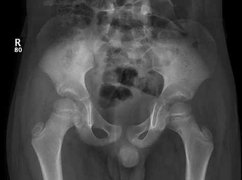
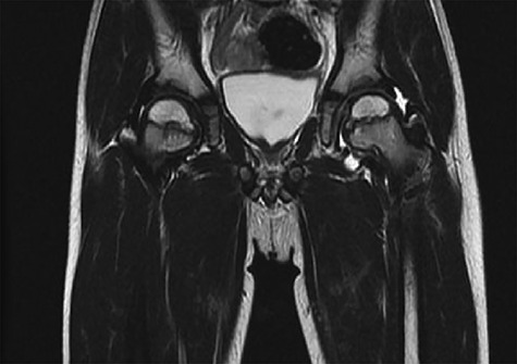
Herniation pits are small benign oval lesions that were reported to be always lying within the super-lateral femoral neck, and were first described in 1982 by Michael J. Pitt. They are usually a unilateral incidental finding along with asymptomatic course. It was widely believed that herniation pits are a result of invagination of the overlying synovium into small cortical defects in the femoral neck. In our case; the mentioned lesions were found atypically bilaterally at the inferomedial aspect of the neck of femur of a 7-year old child. Radiological scans were efficient to obtain an adequate diagnosis whereas conservative management proved to be sufficient dealing with the lesions. Synovial pits may have atypical clinical and radiological course, and this can raise concerns especially with symptomatic hip that may encourage surgical interventions. However, due to benign course of these lesions, we do not recommend any surgical intervention for such lesions.
Keywords: Synovial herniation pits; cortical defect; herniation pits.
Published by Oxford University Press and JSCR Publishing Ltd. All rights reserved. © The Author(s) 2020.
Figures


References
-
- Allen H. A system of human anatomy including its medical and surgical relations, Section II – Bones and joints. Philadelphia, PA: Henry C. Lea’s Sons, 1882, 189–93.
-
- Kate BR. The incidence and cause of cervical fossa in indian femora. J Anat Soc India 1963;12:69–76.
-
- Angel JL. The reaction area of the femoral neck. Clin Orthop 1964;32:89–142. - PubMed
-
- Pitt MJ, Graham AR, Shipman JH, Birkby W. Herniation pit of the femoral neck. AJR Am J Roentgenol 1982;138:1115–21. - PubMed
-
- Leunig M, Beck M, Kalhor M, Kim YJ, Werlen S, Ganz R. Fibrocystic changes at anterosuperior femoral neck: prevalence in hips with femoroacetabular impingement. Radiology 2005;236:237–46. - PubMed
Publication types
LinkOut - more resources
Full Text Sources
