. 2020 Nov 3;30(12):6325-6335.
doi: 10.1093/cercor/bhaa188.

Functional Organization for Response Inhibition in the Right Inferior Frontal Cortex of Individual Human Brains

Affiliations

Functional Organization for Response Inhibition in the Right Inferior Frontal Cortex of Individual Human Brains

Akimitsu Suda et al. Cereb Cortex. .

Abstract

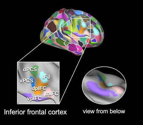
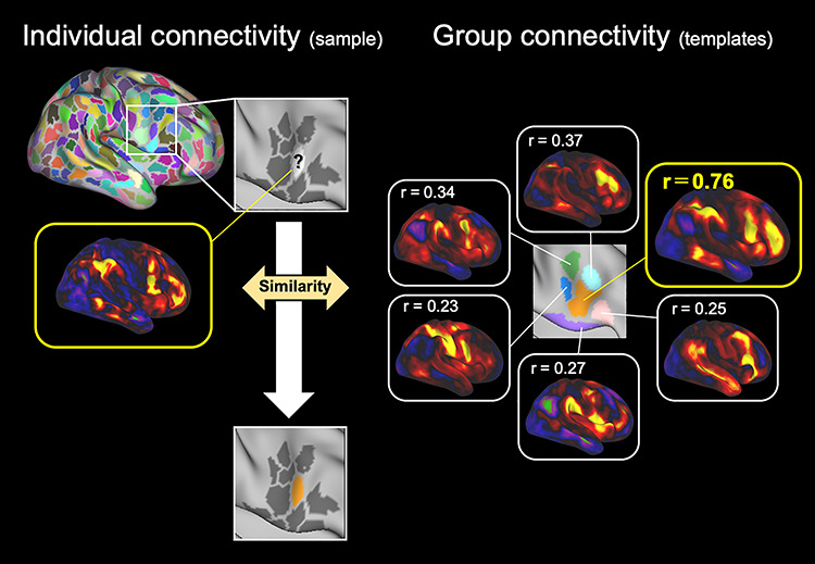
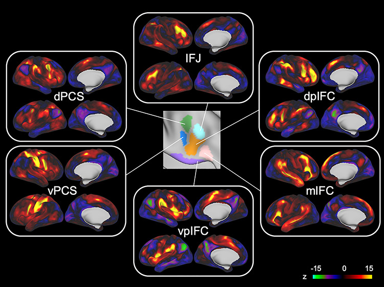
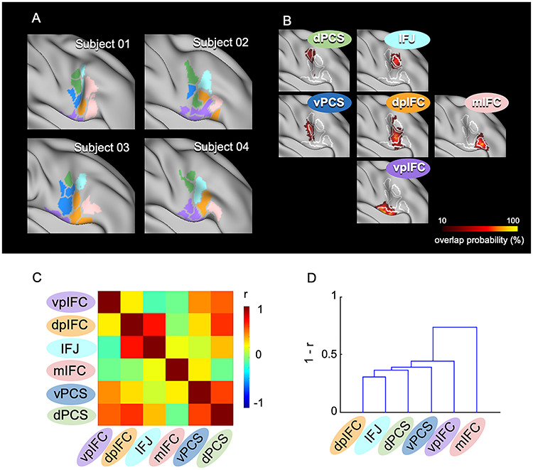
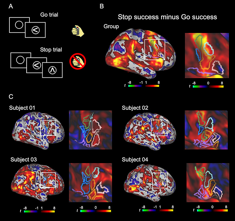
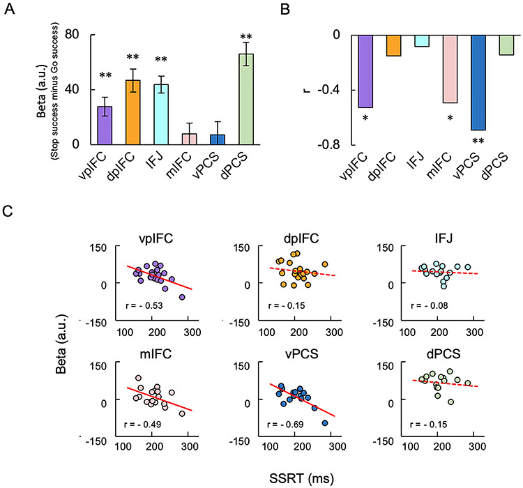
The right inferior frontal cortex (IFC) is critical to response inhibition. The right IFC referred in the human studies of response inhibition is located in the posterior part of the inferior frontal gyrus and the surrounding regions and consists of multiple areas that implement distinct functions. Recent studies using resting-state functional connectivity have parcellated the cerebral cortex and revealed across-subject variability of parcel-based cerebrocortical networks. However, how the right IFC of individual brains is functionally organized and what functional properties the IFC parcels possess regarding response inhibition remain elusive. In the present functional magnetic resonance imaging study, precision functional mapping of individual human brains was adopted to the parcels in the right IFC to evaluate their functional properties related to response inhibition. The right IFC consisted of six modules or subsets of subregions, and the spatial organization of the modules varied considerably across subjects. Each module revealed unique characteristics of brain activity and its correlation to behavior related to response inhibition. These results provide updated functional features of the IFC and demonstrate the importance of individual-focused approaches in studying response inhibition in the right IFC.

Keywords: areal parcellation; boundary mapping; functional connectivity; inferior frontal gyrus; stop-signal task.


Figures

Figure 1
Figure 1
Parcels in the right IFC in group data. There were six parcels in the right IFC in the group data: the vpIFC, shown in purple; dpIFC, shown in orange; IFJ, shown in light blue; mIFC, shown in pink; vPCS, shown in blue; and dPCS, shown in green. These parcels were located in the pars opercularis, which has been shown to be critical to response inhibition, and its surrounding regions in the frontal cortex (the pars triangularis, IFJ, and precentral sulcus).
Figure 2
Figure 2
Assignment of IFC parcels of individual brains to one of six modules. The parcels in the right IFC of individual subjects were assigned to the six modules based on the spatial similarity between the cortical correlation maps for the IFC parcels of individual subjects (left) and the six cortical correlation maps in the group data (right). The dpIFC, shown in orange, had the most similar connectivity pattern, shown in a yellow line.
Figure 3
Figure 3
Cortical correlation maps for six modules in the group data. The cortical correlation maps were generated by calculating the functional connectivity between each of the six parcels in the right IFC and the whole cerebral cortex. The six parcels had distinct connectivity patterns with the cerebral cortex and were used for subsequent analyses to assign the IFC parcels of individual brains.
Figure 4
Figure 4
IFC parcels of individual brains assigned to six modules. (A) The right IFC parcels were assigned to the six modules in four representative subjects. The colors in the parcels indicate the six modules shown in Fig. 1, and the gray parcels indicate parcels not assigned to any of the six modules. (B) Vertex-wise probabilistic maps of the six modules in the right IFC averaged across subjects. (C) The matrix of similarity of the cortical correlation maps among the six modules, averaged across subjects. The color scale indicates the correlation coefficient. (D) The dendrogram of the similarity matrix among the six modules.
Figure 5
Figure 5
The vertex-wise brain activity during response inhibition. (A) The basic structure of the stop-signal task. (B, C) Vertex-wise brain activity maps in the group (B) and in the four representative subjects (C) to inspect the spatial extent of the brain activity. A general linear model was applied to each vertex, and Stop success and Go success trials were contrasted. The color scales indicate t values in the vertices.
Figure 6
Figure 6
Brain activity and brain-behavior correlation in six modules during response inhibition. (A) The brain activity in the six modules in the right IFC averaged across subjects. Error bars indicate the standard error of means of the subjects. **P < 0.01, one-sample t-test. (B) The correlation between the brain activity and SSRT in the six modules. *P < 0.05; **P < 0.01. (C) The scatter plots of the correlation in the six modules.

References

    1. Andersson JL, Skare S, Ashburner J. 2003. How to correct susceptibility distortions in spin-echo echo-planar images: application to diffusion tensor imaging. NeuroImage. 20:870–888. - PubMed
    1. Aron AR, Behrens TE, Smith S, Frank MJ, Poldrack RA. 2007. Triangulating a cognitive control network using diffusion-weighted magnetic resonance imaging (MRI) and functional MRI. J Neurosci. 27:3743–3752. - PMC - PubMed
    1. Aron AR, Fletcher PC, Bullmore ET, Sahakian BJ, Robbins TW. 2003. Stop-signal inhibition disrupted by damage to right inferior frontal gyrus in humans. Nat Neurosci. 6:115–116. - PubMed
    1. Aron AR, Monsell S, Sahakian BJ, Robbins TW. 2004a. A componential analysis of task-switching deficits associated with lesions of left and right frontal cortex. Brain. 127:1561–1573. - PubMed
    1. Aron AR, Poldrack RA. 2006. Cortical and subcortical contributions to stop signal response inhibition: role of the subthalamic nucleus. J Neurosci. 26:2424–2433. - PMC - PubMed

Publication types