

Sex Differences in Anatomic Features Linked to Anterior Cruciate Ligament Injuries During Skeletal Growth and Maturation
- PMID: 32667272
- PMCID: PMC7856525
- DOI: 10.1177/0363546520931831
Sex Differences in Anatomic Features Linked to Anterior Cruciate Ligament Injuries During Skeletal Growth and Maturation
Abstract
Background: Several anatomic features of the knee have been shown to affect joint and anterior cruciate ligament (ACL) loading and the risk of subsequent injuries. While several studies have highlighted sex differences between these anatomic features, little is known on how these differences develop during skeletal growth and maturation.
Hypotheses: (A) Anatomic features linked to an ACL injury will significantly change during skeletal growth and maturation. (B) The age-related changes in anatomic features linked to an ACL injury are different between male and female patients.
Study design: Cross-sectional study; Level of evidence, 3.
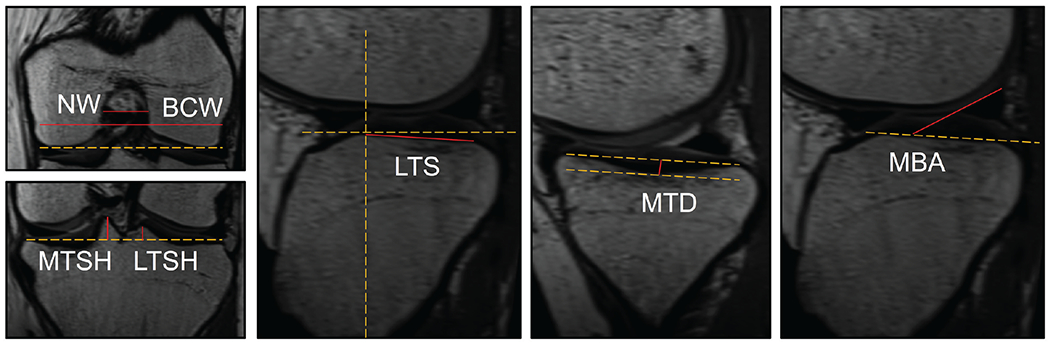
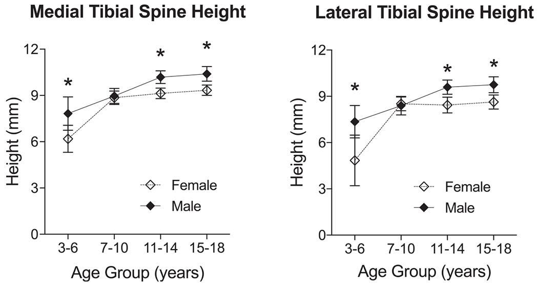
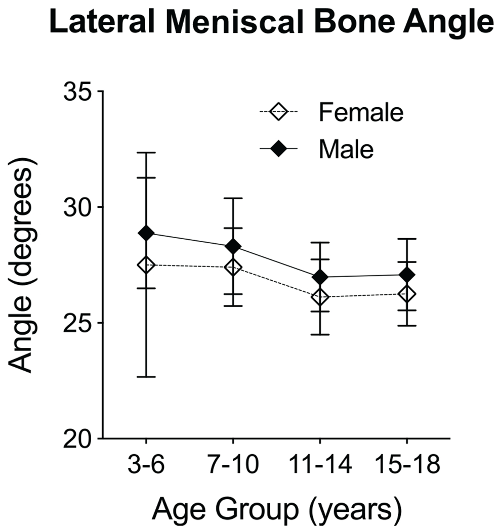
Methods: After institutional review board approval, magnetic resonance imaging data from 269 unique knees (patient age 3-18 years; 51% female), free from any injuries, were used to measure femoral notch width, posterior slope of the lateral tibial plateau (lateral tibial slope), medial tibial depth, tibial spine height, and posterior lateral meniscal bone angle. Linear regression was used to test the associations between age and quantified anatomic indices. Patients were then divided into 4 age groups: preschool (3-6 years), prepubertal (7-10 years), early adolescent (11-14 years), and late adolescent (15-18 years). Also, 2-way analysis of variance with the Holm-Sidak post hoc test was used to compare morphology between male and female patients in each age group.
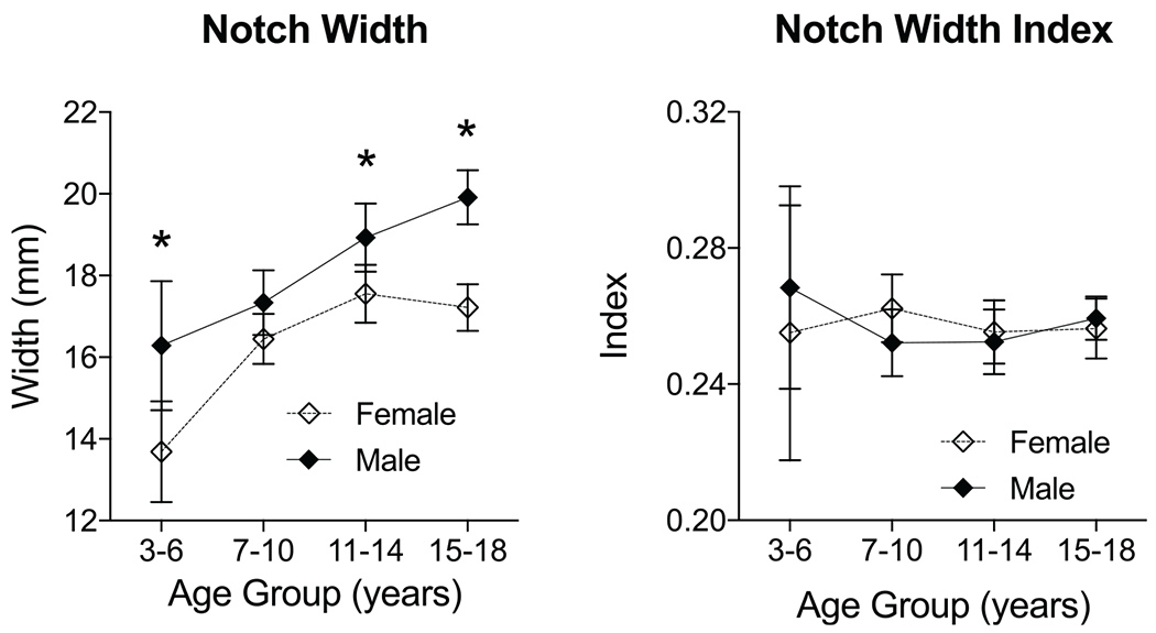
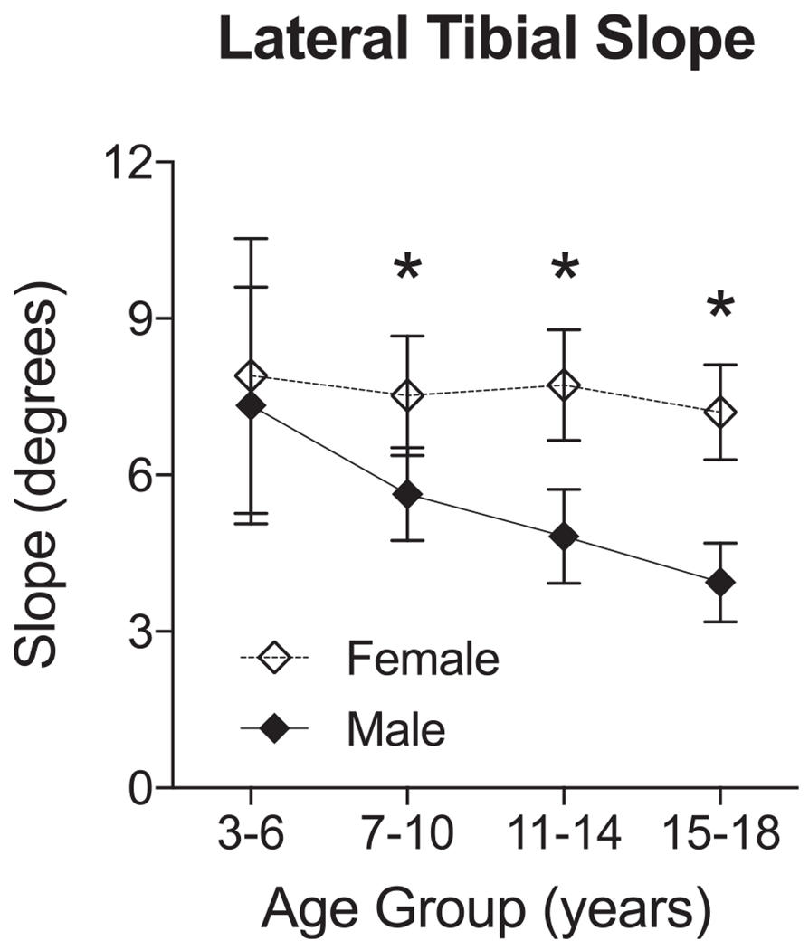
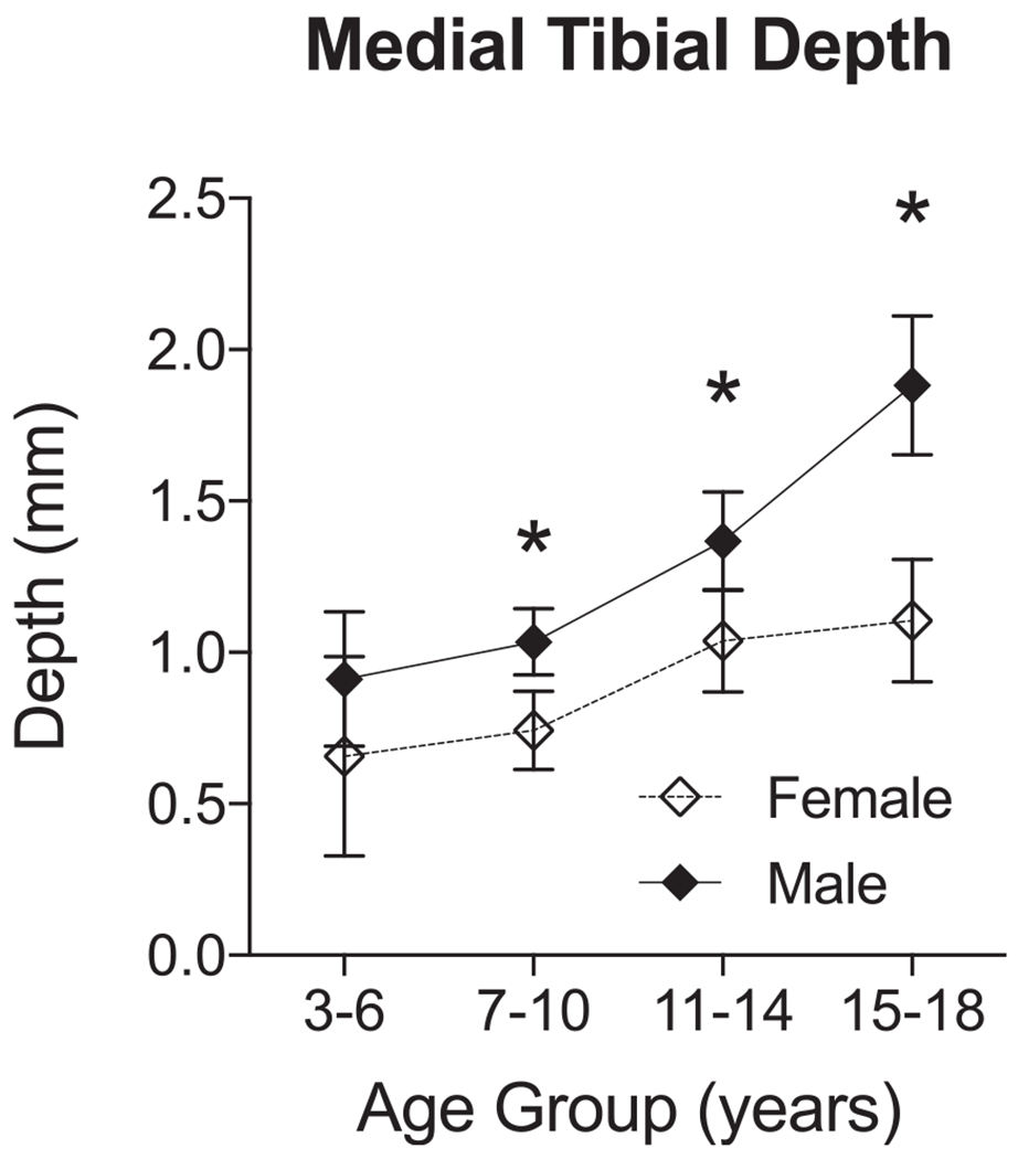
Results: The femoral notch width, medial tibial depth, and tibial spine height significantly increased with age (P < .001). The lateral tibial slope decreased with age only in male patients (P < .001). Except for the posterior lateral meniscal bone angle, the age-related changes in anatomy were different between male and female patients (P < .05). On average, early and late adolescent female patients had smaller femoral notches, steeper lateral tibial slopes, flatter medial tibial plateaus, and shorter tibial spines compared with age-matched male patients (P < .01).
Conclusion: Overall, the findings supported our hypotheses, showing sex-specific changes in anatomic features linked to an ACL injury during skeletal growth and maturation. These observations help to better explain the reported age and sex differences in the prevalence of ACL injuries. The fact that most of these anatomic features undergo substantial changes during skeletal growth and maturation introduces the hypothesis that prophylactic interventions (ie, activity modification) would have the potential to reshape a maturing knee in a manner that lowers the risk of noncontact ACL injuries.
Keywords: ACL; anatomy; knee; sex; skeletal maturation.
Figures
References
-
- Ageberg E, Forssblad M, Herbertsson P, Roos EM. Sex differences in patient-reported outcomes after anterior cruciate ligament reconstruction: data from the Swedish knee ligament register. Am J Sports Med. 2010;38(7):1334–1342. - PubMed
-
- Anderson AF, Dome DC, Gautam S, Awh MH, Rennirt GW. Correlation of anthropometric measurements, strength, anterior cruciate ligament size, and intercondylar notch characteristics to sex differences in anterior cruciate ligament tear rates. Am J Sports Med. 2001;29(1):58–66. - PubMed
-
- Beynnon BD, Hall JS, Sturnick DR, et al. Increased slope of the lateral tibial plateau subchondral bone is associated with greater risk of noncontact ACL injury in females but not in males: a prospective cohort study with a nested, matched case-control analysis. Am J Sports Med. 2014;42(5):1039–1048. - PMC - PubMed
-
- Cavaignac E, Perroncel G, Thepaut M, Vial J, Accadbled F, De Gauzy JS. Relationship between tibial spine size and the occurrence of osteochondritis dissecans: an argument in favour of the impingement theory. Knee Surg Sports Traumatol Arthrosc. 2017;25(8):2442–2446. - PubMed
Publication types
MeSH terms
Grants and funding
LinkOut - more resources
Full Text Sources
