Clinical Trial
. 2020 Jul 15:26:e924593.
doi: 10.12659/MSM.924593.

Argatroban Increased the Basal Vein Drainage and Improved Outcomes in Acute Paraventricular Ischemic Stroke Patients

Affiliations
Clinical Trial

Argatroban Increased the Basal Vein Drainage and Improved Outcomes in Acute Paraventricular Ischemic Stroke Patients

Shoufeng Liu et al. Med Sci Monit. .

Abstract

BACKGROUND Since venous drainage in acute arterial ischemic stroke has not been thoroughly researched, we evaluate the effect of argatroban, a selective direct thrombin inhibitor, as a therapy to increase the rate of basal vein Rosenthal (BVR) drainage and improve patients' post-stroke outcomes. MATERIAL AND METHODS In this multicenter clinical trial, 60 eligible patients at 4.5 to 48 hours after the stroke onset were recruited. After being randomly allocated into 2 groups, they were treated with standard therapy either alone or with argatroban. RESULTS Compared to the contralateral brain hemisphere, the mean flow velocity (MFV) in BVR drainage was significantly reduced in the stroke-afflicted ipsilateral hemisphere. After treatment with argatroban for 7 days, the MFV from BVR of the ipsilateral hemisphere in the argatroban treated group was significantly increased when compared to the control group. At 90 days after the onset of stroke, the MFV of BVR in the ipsilateral hemisphere was similar in both groups. Compared with controls, the argatroban-treated patients had smaller lesions from baseline to 7 days. Argatroban also improved National Institutes of Health Stroke Scale (NIHSS) scores on day 7 after the onset of stroke. Furthermore, the argatroban group's neurological functions were superior to those of their untreated counterparts after 90 days. No difference was found in the incidence of adverse reactions between the 2 groups. CONCLUSIONS These observations indicate that vein drainage change may contribute to the acute phase of brain edema and the outcomes of ischemic stroke patients.


Conflict of interest statement

Conflict of interest

None.

Figures

Figure 1
Figure 1
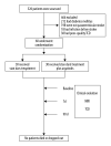
Effect of argatroban on BVR drainage in acute ischemic stroke: trial profile. Sixty patients with acute paraventricular ischemic stroke and matched clinical characteristics, who were beyond the 4.5-hour window for alteplase when recruited, were randomized into 2 groups. Both groups received standard stroke therapy, and one group (n=30) also received 10 mg intravenous argatroban (twice daily for 7 days consecutively). Treatment began between 1 hour after a baseline MRI and 48 hours after symptom onset. NIHSS, mBI, and mRS were conducted. Lesion volume was measured by MRI on admission. MFV in the BVR was measured by TCD at baseline and on day 7 and day 90. BVR – basal vein Rosenthal; MRI – magnetic resonance imaging; NIHSS – National Institutes of Health Stroke Scale; MFV – mean flow velocity; TCD – transcranial Doppler ultrasonography.
Figure 2
Figure 2
MFV in bilateral BVR of the patients with acute paraventricular ischemic stroke at baseline and after argatroban therapy. (A) Comparison of MFV in ipsilateral versus contralateral BVR between control and argatroban-treated patients. (B) Impact of argatroban on venous flow in the BVR of patients’ ipsilateral hemisphere. (C) Impact of argatroban on venous flow in BVR of patients’ contralateral hemisphere. (D) Representative TCD illustrates the MFV in BVRs of ipsilateral hemispheres in patients at the baseline and at day 7 and day 90 after argatroban treatment. # P<0.05 versus contralateral hemisphere at baseline, * P<0.05, ** P<0.05 compared to baseline of argatroban group, *** P<0.05 compared to control at the same time point. BVR – basal vein Rosenthal; MFV – mean flow velocity; TCD – transcranial Doppler ultrasonography.
Figure 3
Figure 3
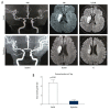
Impact of argatroban on lesion volume growth in patients. (A) MRI images show acute right hemisphere infarct with middle cerebral artery stenosis in a control patient (top) and acute right hemisphere infarct with middle cerebral artery stenosis in an argatroban-treated patient (bottom). Lesion volumes were measured on a DWI (baseline) and T2FLAIR (day 7). (B) Growth of lesion volume from baseline to day 7. Lesion growth equals lesion volume measured on T2FLAIR at day 7 minus that measured on DWI at baseline. Data are presented as mean±SD. An independent t-test was conducted for each comparison. MRI – magnetic resonance imaging; DWI – diffusion weighted imaging; SD – standard deviation.
Figure 4
Figure 4
Impact of argatroban on clinical outcomes in the argatroban group compared to the control group. (A) Trends of NIHSS scores from argatroban-treated and control patients. (B) NIHSS values for argatroban-treated and control patients in the 7 days (NIHSS change=baseline – day 7). (C) Comparison of mBI between groups. (D) mRS difference between groups at day 90. * P<0.05 compared to the control at the same time point. NIHSS – National Institutes of Health Stroke Scale; mBI – modified Barthel Index; mRS – modified Rankin Scale.

References

    1. Chwojnicki K, Ryglewicz D, Wojtyniak B, et al. Acute ischemic stroke hospital admissions, treatment, and outcomes in Poland in 2009–2013. Front Neurol. 2018;9:134. - PMC - PubMed
    1. Zhang JH, Obenaus A, Liebeskind DS, et al. Recanalization, reperfusion, and recirculation in stroke. J Cereb Blood Flow Metab. 2017;37(12):3818–23. - PMC - PubMed
    1. Manning NW, Warne CD, Meyers PM. Reperfusion and clinical outcomes in acute ischemic stroke: systematic review and meta-analysis of the Stent-Retriever-Based, Early Window Endovascular Stroke Trials. Front Neurol. 2018;9:301. - PMC - PubMed
    1. Iglesias-Rey R, Rodríguez-Yáñez M, Rodríguez-Castro E, et al. Worse outcome in stroke patients treated with rt-PA without early reperfusion: Associated factors. Transl Stroke Res. 2018;9(4):347–55. - PMC - PubMed
    1. Puetz V, Gerber JC, Krüger P, et al. Cerebral venous drainage in patients with space-occupying middle cerebral artery infarction: Effects on functional outcome after hemicraniectomy. Front Neurol. 2018;9:876. - PMC - PubMed

MeSH terms