. 2020 Sep 3;79(5):812-823.e4.
doi: 10.1016/j.molcel.2020.06.031. Epub 2020 Jul 14.

Structural Insights of Transcriptionally Active, Full-Length Androgen Receptor Coactivator Complexes

Affiliations

Structural Insights of Transcriptionally Active, Full-Length Androgen Receptor Coactivator Complexes

Xinzhe Yu et al. Mol Cell. .

Abstract

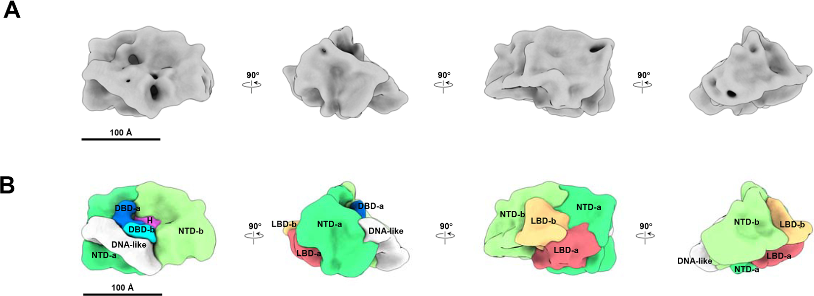
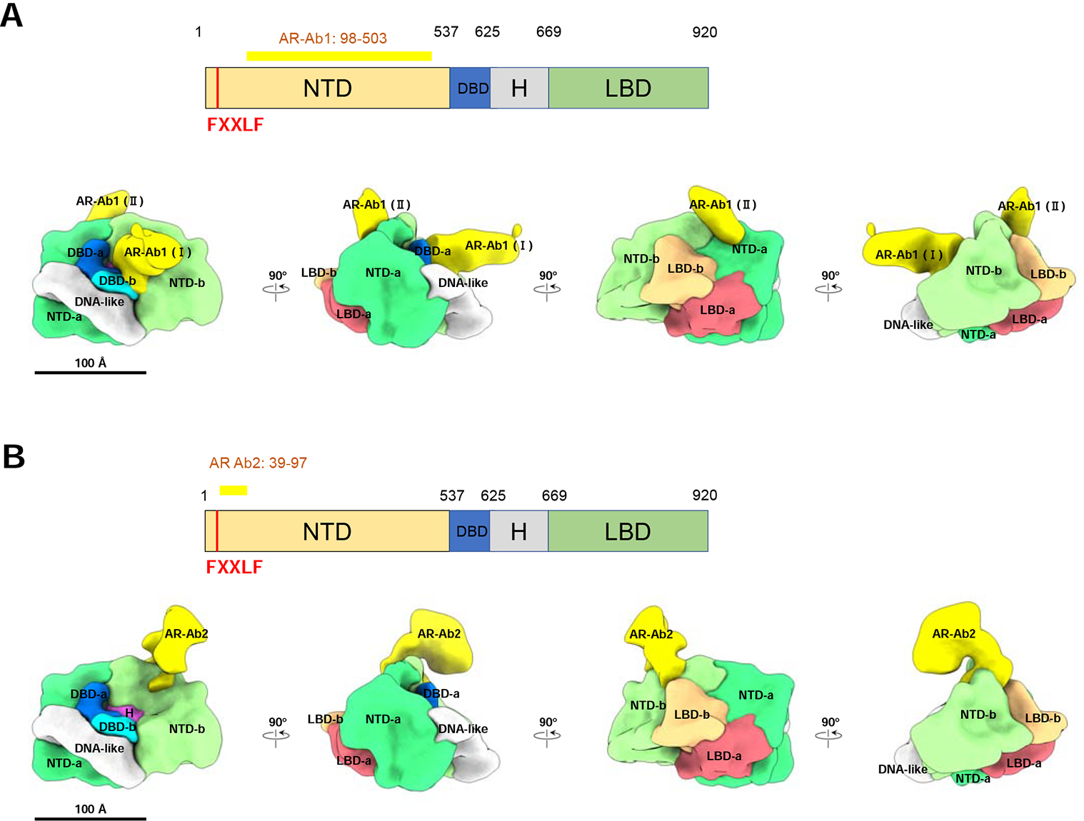
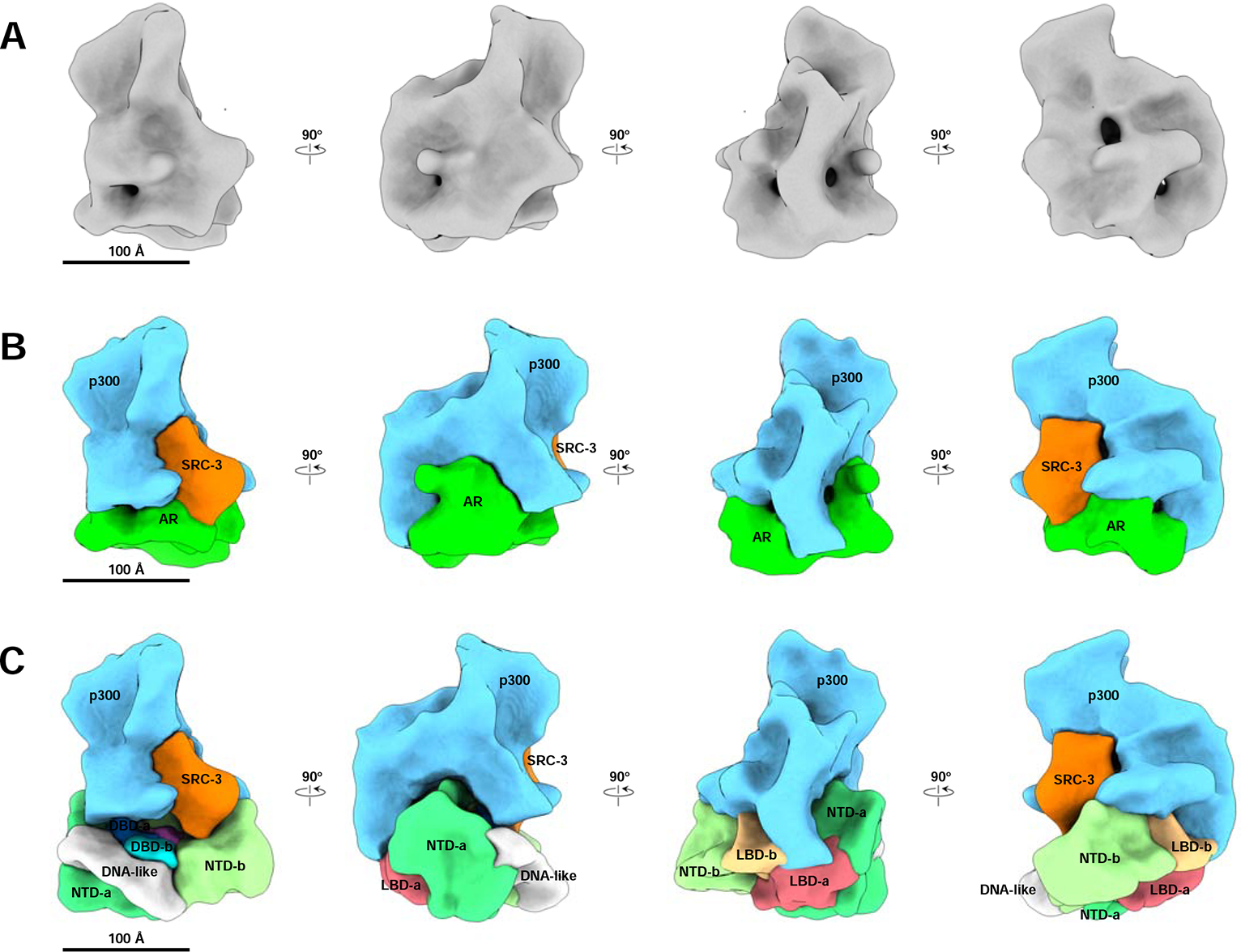
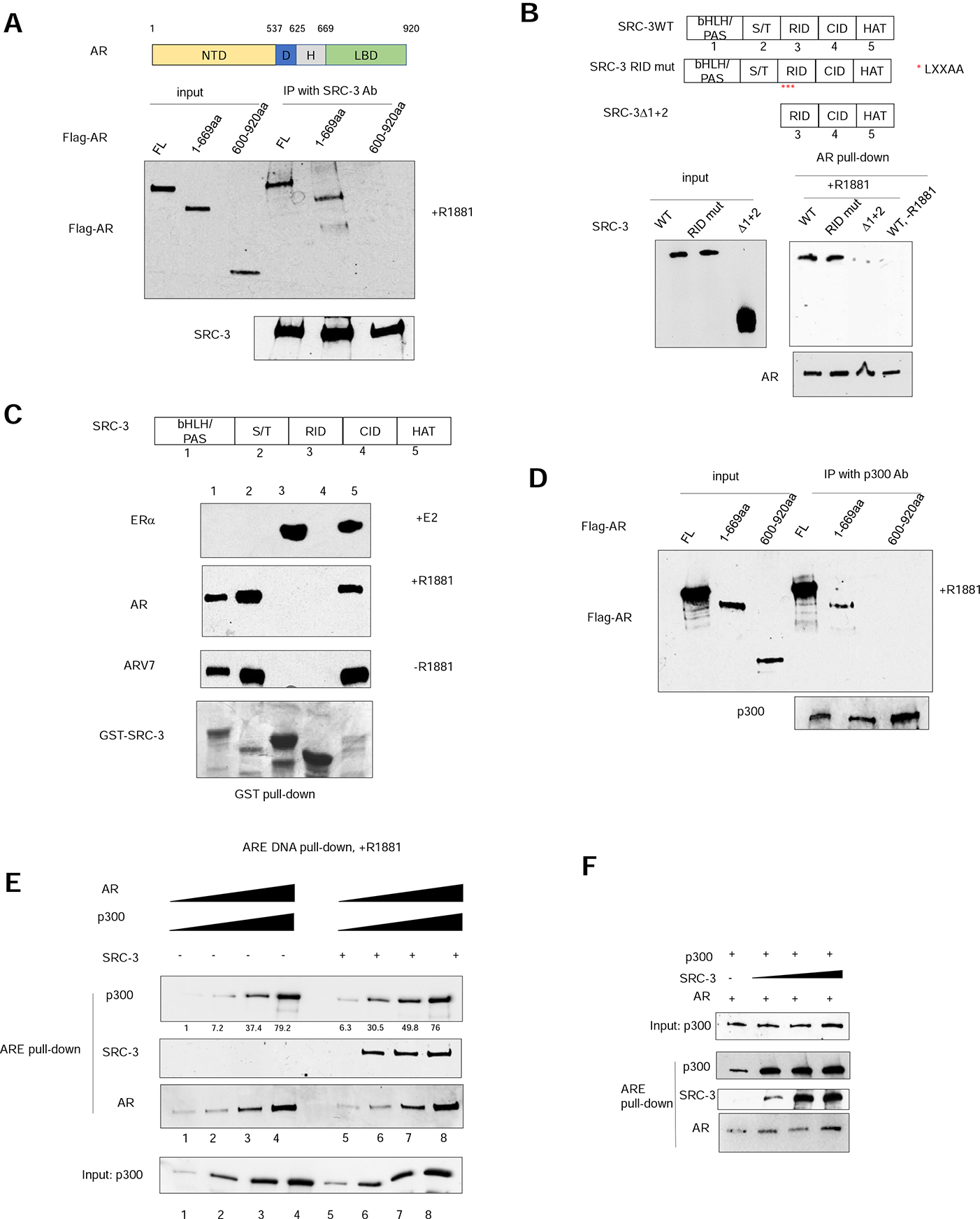
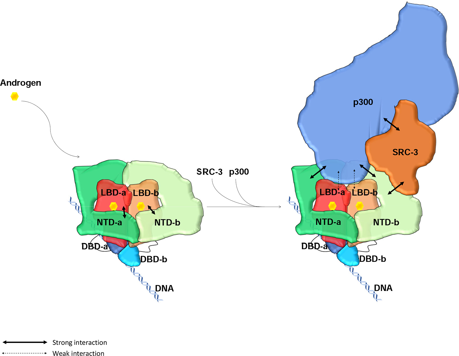
Steroid receptors activate gene transcription by recruiting coactivators to initiate transcription of their target genes. For most nuclear receptors, the ligand-dependent activation function domain-2 (AF-2) is a primary contributor to the nuclear receptor (NR) transcriptional activity. In contrast to other steroid receptors, such as ERα, the activation function of androgen receptor (AR) is largely dependent on its ligand-independent AF-1 located in its N-terminal domain (NTD). It remains unclear why AR utilizes a different AF domain from other receptors despite that NRs share similar domain organizations. Here, we present cryoelectron microscopy (cryo-EM) structures of DNA-bound full-length AR and its complex structure with key coactivators, SRC-3 and p300. AR dimerization follows a unique head-to-head and tail-to-tail manner. Unlike ERα, AR directly contacts a single SRC-3 and p300. The AR NTD is the primary site for coactivator recruitment. The structures provide a basis for understanding assembly of the AR:coactivator complex and its domain contributions for coactivator assembly and transcriptional regulation.

Keywords: AR dimerization; N-terminal domain; N/C interaction; SRC-3; androgen receptor; coactivator; complex; cryo-EM; p300; structure.


Conflict of interest statement

Declaration of Interests The authors declare no competing interests.

Figures

Figure 1.
Figure 1.. ARE DNA bound AR density map and segmentation.
(A) Cryo-EM density of ARE-DNA/AR at resolution ~12.6 Å viewed from different orientation rotating in vertical direction. (B) Segmentation result of ARE-DNA/AR. The segments of two ARs are shown as -a and -b separately. The functional domains of AR are annotated in different colors. NTD: N-terminal domain (Green and light Green), LBD: ligand binding domain (Orange and Red), DBD: DNA binding domain (Blue and light Blue), H: the Hinge region (Purple) between DBD and LBD and one DNA-like feature is shown in White. See also Supplemental Figure 1 and Supplemental Table 1.
Figure 2.
Figure 2.. NTD and LBD annotation validation by antibody labeling.
(A) AR-Ab1 labels the NTDs. Top panel, schematic representation of AR functional domains. The long yellow bar represents the region (residues 98–503) recognized by the AR-Ab1. The vertical red bar represents the FXXLF motif (residue 23–27). Bottom panel, AR-Ab1 (yellow) binding to the NTD-a and -b in segmented ARE-DNA/AR density. The stronger antibody density is labeled as AR-Ab1 (Ⅰ) and the other one is labeled as AR-Ab1 (Ⅱ). (B) AR-Ab2 labels the NTD and the interface between the NTD and LBD. Top panel, AR-Ab2 recognizes the residues 39–97 (horizontal yellow bar) at the N-terminal end of AR, which is adjacent to the FXXLF, a motif mediating the interaction between NTD and LBD. Bottom panel, the AR-Ab2 (yellow) binding to the NTD-b in segmented ARE-DNA/AR density. It is located close to the interface between NTD-b and LBD-b, consistent with the segmentation result. See also Supplemental Figure 2.
Figure 3.
Figure 3.. The ARE DNA-bound AR/SRC-3/p300 complex density map and segmentation.
(A) Cryo-EM density map of the ARE DNA-bound AR/SRC-3/p300 complex at resolution ~20 Å. Shown are 4 different angles of the map rotating every 90 degrees. (B) Segmentation of ARE-DNA/AR/SRC-3/p300. Each component was segmented to annotate different proteins: AR, Green; SRC-3, Orange; p300, Blue, respectively. (C) The assembled ARE-DNA/AR/SRC-3/p300 structure with segmented ARE-DNA/AR density (Figure 1B) replacing the AR density. The p300 density mainly interacts with two NTDs of AR and has a small area touches the two LBDs. See also Supplemental Figure 3, 4, 5 and Supplemental Table 1.
Figure 4.
Figure 4.. The AR NTD is essential for SRC-3 and p300 coactivator recruitment.
(A) AR interacts with SRC-3 through its N-terminal domain (NTD) in vitro. Purified flag-tagged full-length AR, AR(1–669aa) or AR (600–920aa) was incubated with purified, recombinant SRC-3 in the presence of 1 μM R1881 followed by co-immunoprecipitation using an SRC-3-specific antibody. (B) Deletion of two N-terminal domains of SRC-3, but not RID LXXAA mutations, abolished AR-SRC-3 interaction in vitro. Flag-tagged SRC-3 wild-type (WT) or mutant was transiently transfected into 293T cells and the lysates were incubated with purified, recombinant AR protein in the absence or presence of 1μM R1881 followed by co-immunoprecipitation using an AR-specific antibody. Red asterisks represent mutation of the three LXXLL motifs present in SRC-3 RID into LXXAA. (C) The SRC-3 RID does not interact with AR in a GST pull-down assay. Glutathione-sepharose beads bound to different GST-SRC-3 fragments were incubated with purified ERα, AR or ARV7 protein in the absence or presence of corresponding ligand. Top 3 panels, Immunoblotting analysis of glutathione bead-bound receptor proteins. Bottom panel, Coomassie blue staining of the different tested GST-SRC-3 fragments. (D) AR interacts with p300 through its N-terminal domain in vitro. Purified flag-tagged full-length AR, AR (1–669aa) or AR (600–920aa) was incubated with purified, recombinant p300 in the presence of 1 μM R1881 followed by co-immunoprecipitation using a p300-specific antibody. (E) SRC-3 increases the interaction between AR and p300 when both are at low concentrations. A series of concentrations of purified AR, p300, ARE-containing biotinylated DNA were incubated with or without purified SRC-3 in the presence of 1μM R1881 followed by magnetic streptavidin beads pull-down. Quantitation of p300 pulled-down by ARE DNA is provided underneath the top panel. (F) The effect of different concentrations of purified SRC-3 on the interaction between p300 and AR at low concentrations. Purified p300 protein was incubated with 1μM R1881, AR, and ARE DNA in the absence or presence of increasing concentrations of SRC-3 protein followed by magnetic streptavidin bead pull-down. See also Supplemental Figure 4.
Figure 5:
Figure 5:. Model of full-length AR domain organization and its NTD-mediated SRC-3 and p300 coactivator recruitment.
AR dimerizes upon binding to its ligand androgen. Its LBD and DBD are located at the center to form a tight dimerization interface. The two NTDs in the dimer adopt slightly different conformations, wrap around the LBDs, and make tight intra- and inter-molecular N/C interactions. The NTDs also connect to each other to contribute to the dimerization. Through these interactions, AR dimer forms a unique head-to-head and tail-to-tail dimer. AR dimer then recruits key coactivators, SRC-3 and p300, to ARE DNA mainly through its NTDs. One molecule of SRC-3 interacts with one of the AR NTDs (NTD-b) while p300 interacting with both AR NTDs as well as the LBDs to form a stable transcriptionally active complex.

References

    1. Alen P, Claessens F, Verhoeven G, Rombauts W, and Peeters B (1999). The androgen receptor amino-terminal domain plays a key role in p160 coactivator-stimulated gene transcription. Mol Cell Biol 19, 6085–6097. - PMC - PubMed
    1. Antonarakis ES, Lu C, Wang H, Luber B, Nakazawa M, Roeser JC, Chen Y, Mohammad TA, Fedor HL, Lotan TL, et al. (2014). AR-V7 and resistance to enzalutamide and abiraterone in prostate cancer. N Engl J Med 371, 1028–1038. - PMC - PubMed
    1. Balbas MD, Evans MJ, Hosfield DJ, Wongvipat J, Arora VK, Watson PA, Chen Y, Greene GL, Shen Y, and Sawyers CL (2013). Overcoming mutation-based resistance to antiandrogens with rational drug design. Elife 2, e00499. - PMC - PubMed
    1. Bell JM, Chen M, Baldwin PR, and Ludtke SJ (2016). High resolution single particle refinement in EMAN2.1. Methods 100, 25–34. - PMC - PubMed
    1. Brzozowski AM, Pike AC, Dauter Z, Hubbard RE, Bonn T, Engstrom O, Ohman L, Greene GL, Gustafsson JA, and Carlquist M (1997). Molecular basis of agonism and antagonism in the oestrogen receptor. Nature 389, 753–758. - PubMed

Publication types

MeSH terms