. 2020 Jul 14;32(2):107898.
doi: 10.1016/j.celrep.2020.107898.

The Cdc48 Complex Alleviates the Cytotoxicity of Misfolded Proteins by Regulating Ubiquitin Homeostasis

Affiliations

The Cdc48 Complex Alleviates the Cytotoxicity of Misfolded Proteins by Regulating Ubiquitin Homeostasis

Ryan Higgins et al. Cell Rep. .

Abstract

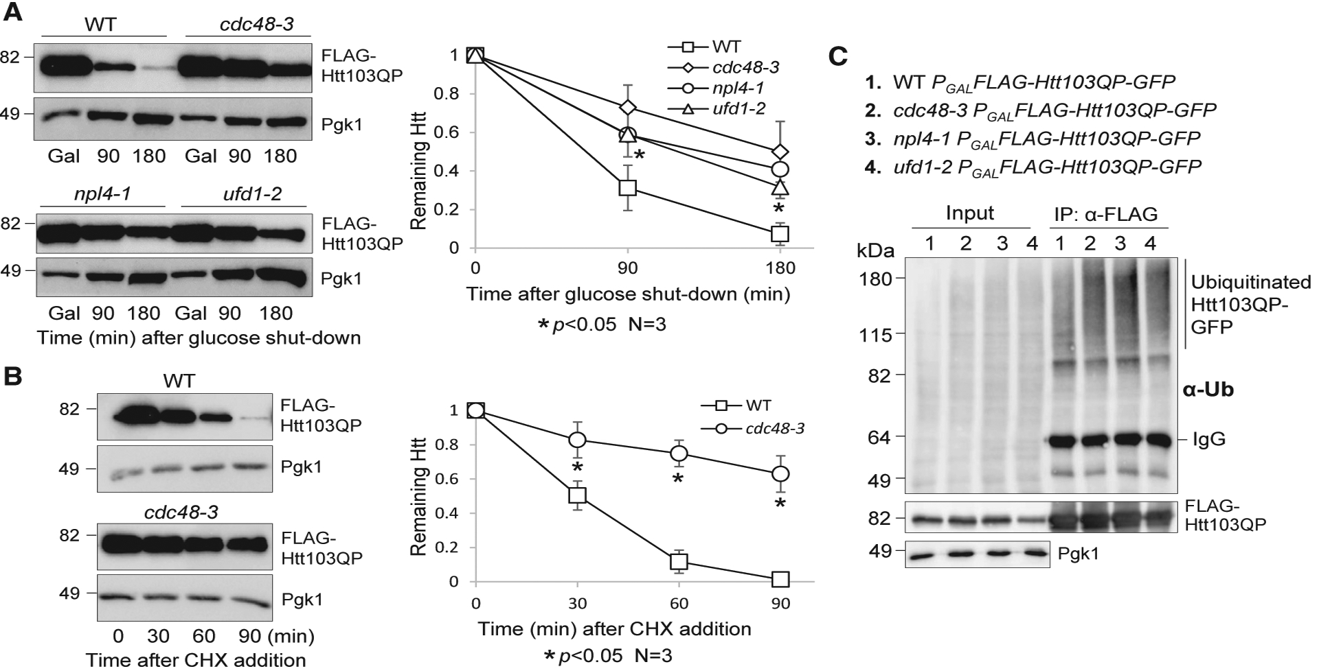
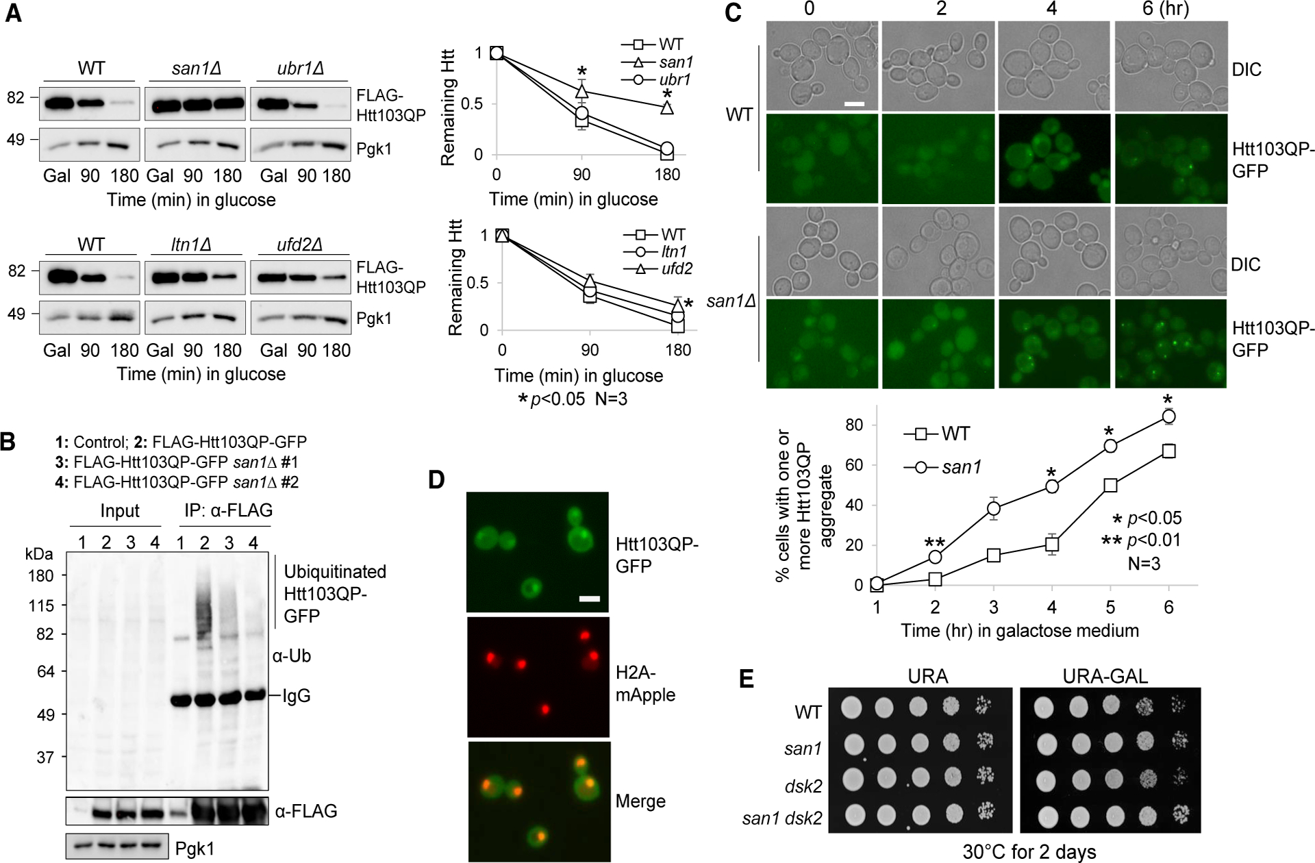
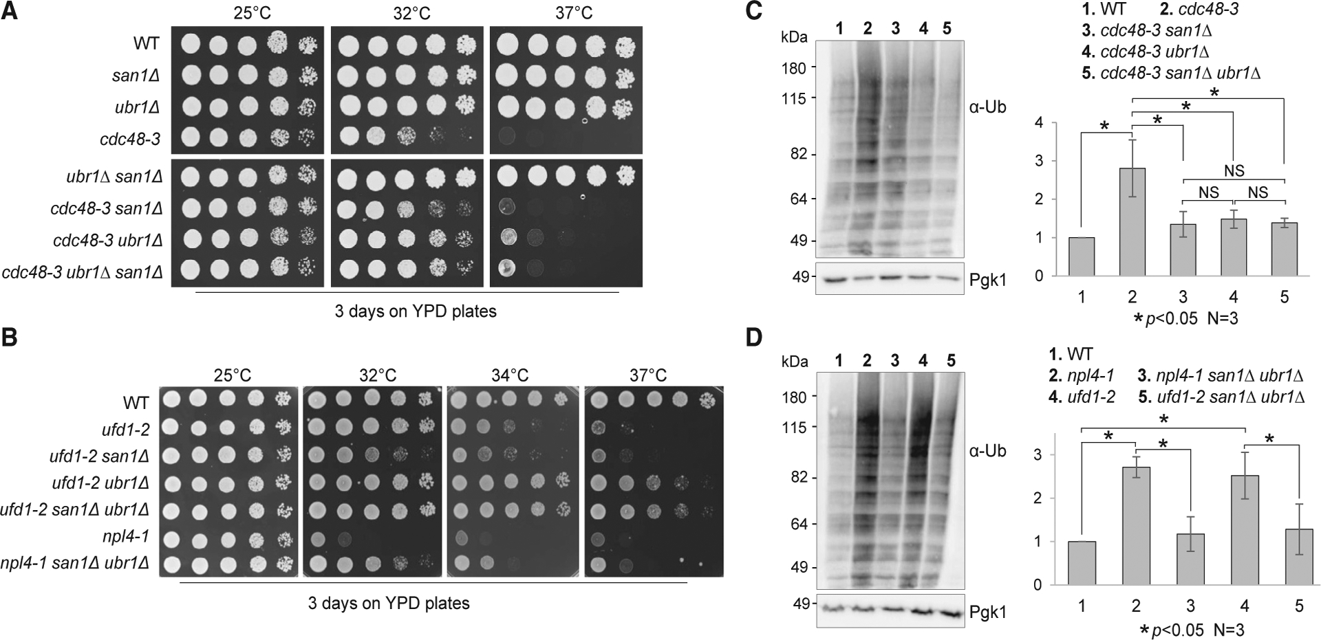
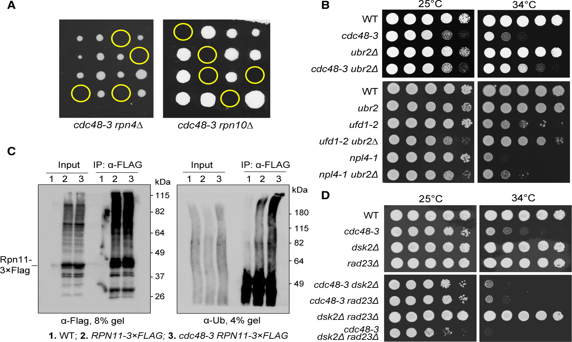
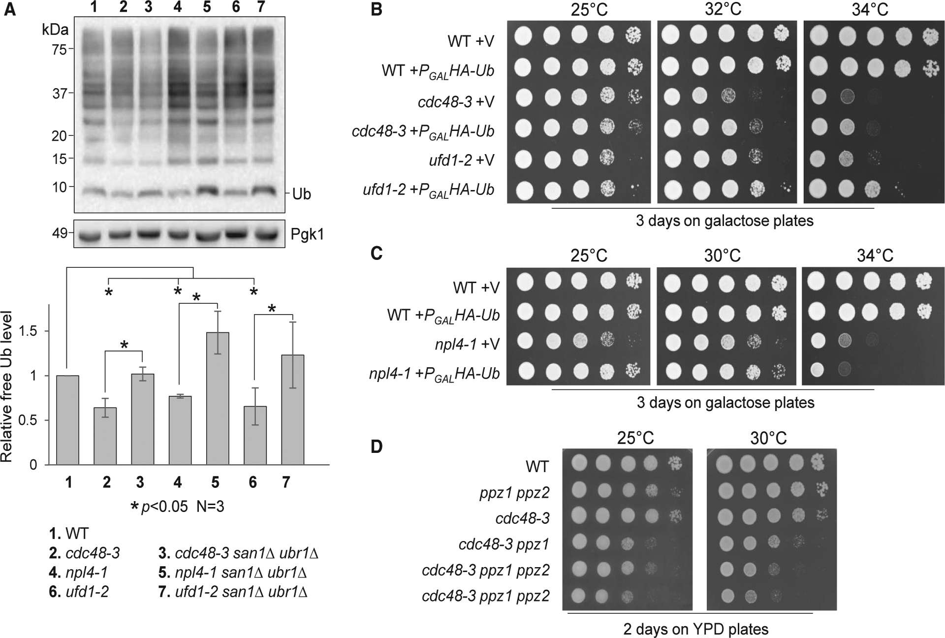
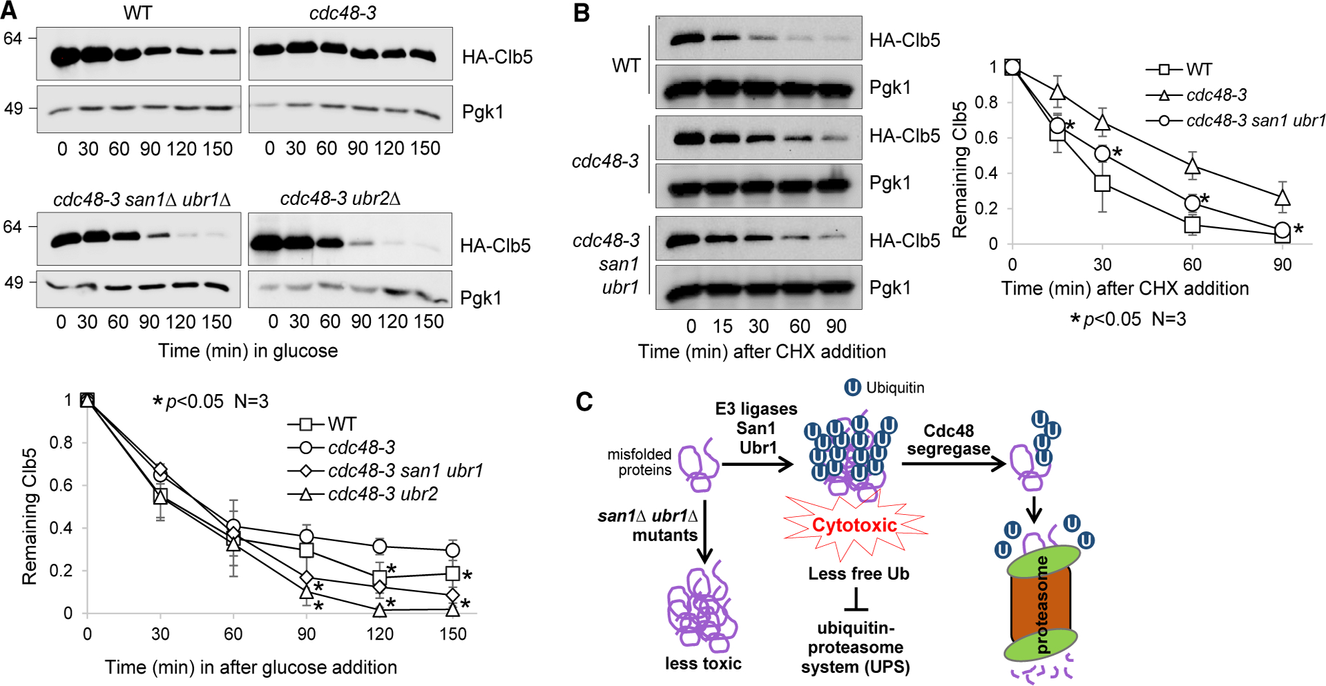
The accumulation of misfolded proteins is associated with multiple neurodegenerative disorders, but it remains poorly defined how this accumulation causes cytotoxicity. Here, we demonstrate that the Cdc48/p97 segregase machinery drives the clearance of ubiquitinated model misfolded protein Huntingtin (Htt103QP) and limits its aggregation. Nuclear ubiquitin ligase San1 acts upstream of Cdc48 to ubiquitinate Htt103QP. Unexpectedly, deletion of SAN1 and/or its cytosolic counterpart UBR1 rescues the toxicity associated with Cdc48 deficiency, suggesting that ubiquitin depletion, rather than compromised proteolysis of misfolded proteins, causes the growth defect in cells with Cdc48 deficiency. Indeed, Cdc48 deficiency leads to elevated protein ubiquitination levels and decreased free ubiquitin, which depends on San1/Ubr1. Furthermore, enhancing free ubiquitin levels rescues the toxicity in various Cdc48 pathway mutants and restores normal turnover of a known Cdc48-independent substrate. Our work highlights a previously unappreciated function for Cdc48 in ensuring the regeneration of monoubiquitin that is critical for normal cellular function.

Keywords: Cdc48; San1/Ubr1 E3 ligases; mutated Huntingtin; proteotoxicity; ubiquitin homeostasis.


Conflict of interest statement

Declaration of Interests There are no competing interests.

Figures

Figure 1.
Figure 1.. The Cdc48Npl4/Ufd1 Complex Is Required for Htt103QP Degradation but Is Dispensable for Its Ubiquitination
(A) The degradation of Htt103QP depends on the Cdc48Npl4/Ufd1 complex. WT (3419–1-1), cdc48–3 (3598–2-3), npl4–1 (3387–3-4), and ufd1–2 (3385–4-4) mutants containing an integrating plasmid, PGALFLAG-Htt103QP-GFP, were grown in non-inducing raffinose medium (YPR) to mid-log phase at 25°C. Galactose was added to the medium (2% final concentration) for 50 min to induce FLAG-Htt103QP-GFP expression at 25°C. Cells were then shifted to 34°C for 10 additional min, and glucose was added to shut off the expression. FLAG-Htt103QP-GFP protein levels were detected using anti-FLAG antibody. Pgk1, loading control. The levels of Htt103QP and Pgk1 are shown on the left. The intensity of protein bands and the Htt103QP/Pgk1 ratio were analyzed using ImageJ. The remaining Htt103QP after glucose shutoff was calculated based on the results from three independent experiments. The results are represented as mean ± SD (standard deviation). Wilcoxon rank-sum test was used to calculate the p values. The statistical difference is significant (*) when p < 0.05. (B) Impaired Htt103QP degradation was estimated by cycloheximide (CHX) chase. WT (3419–1-1) and cdc48–3 (3598–2-3) strains with PGALFLAG-Htt103QP-GFP were grown in YPR at 25°C to mid-log phase. Galactose was added to the medium for 30 min, followed by temperature shift to 34°C for 30 min. Then, CHX (200 μg/mL) was added, and the protein levels of Htt103QP and Pgk1 were determined over time. The experiment was repeated three times. Quantification of the Htt103QP/Pgk1 ratio and statistical analysis were performed as described above, and the relative levels of Htt103QP to Pgk1 are represented as mean ± SD. *p < 0.05. (C) Cdc48 is not required for Htt103QP ubiquitination. WT (3419–1-1), cdc48–3 (3598–2-3), npl4–1 (3387–3-4), and ufd1–2 (3385–4-4) mutant cells containing PGALFLAG-Htt103QP-GFP were grown in YPR to mid-log phase at 25°C. Galactose was then added, and cells were shifted to 34°C for 3 h. The protein extracts were prepared as described in the STAR Methods. FLAG-Htt103QP-GFP was immunoprecipitated using M2 anti-FLAG beads. Htt103QP protein levels were detected using an anti-FLAG antibody, and protein ubiquitination was detected using anti-Ub antibody. Pgk1, loading control.
Figure 2.
Figure 2.. E3 Ubiquitin Ligase San1 Ubiquitinates Htt103QP for Degradation
(A) San1 is required for Htt103QP degradation. WT (3419–1-1), san1Δ (RH142), ubr1Δ (3522–4-4), ltn1Δ (3287–1-1), and ufd2Δ (3288–1-3) cells containing PGALFLAG-Htt103QP-GFP were grown in YPR to mid-log phase at 30°C. Galactose was then added to induce Htt103QP expression for 1 h. Glucose was then added to shut off galactose-induced expression. Htt103QP protein levels were detected using an anti-FLAG antibody. The experiment was repeated three times. The western blotting result for Htt103QP level is shown on the left panel. Pgk1, loading control. Protein band intensity and Htt103QP/Pgk1 ratio were analyzed using ImageJ. Quantification results are represented as mean ± SD (right panel). * indicates statistical significant (p < 0.05). (B) San1 promotes ubiquitination of Htt103QP. WT (3419–1-1) and two san1Δ mutant strains (RH142 and 3301–2-2) containing PGALFLAG-Htt103QP-GFP were grown in YPR to mid-log phase at 30°C. Galactose was then added to induce Htt103QP overexpression for 3 h. After preparation of cell lysates, Htt103QP was immunoprecipitated using M2 anti-FLAG beads. Htt103QP protein levels were detected using anti-FLAG antibody, and the ubiquitination level was detected using anti-Ub antibody. Pgk1, loading control. (C) Htt103QP inclusion bodies form at an accelerated rate in san1Δ mutant. WT (3419–1-1) and san1Δ (RH142) cells containing PGALFLAG-Htt103QP-GFP were grown in YPR to mid-log phase at 30°C. Galactose was then added to induce Htt103QP expression. Differential interference contrast (DIC) and GFP fluorescence images were obtained every h for 6 h. Scale bar, 5 μm. The quantitative result is the average from three independent experiments. Data are represented as mean ± SD. Two-way ANOVA with Tukey’s multiple comparisons test was performed for each dataset. *p < 0.05. (D) Htt103QP shows nuclear localization. Yeast cells with H2A-mApple and PGALFLAG-Htt103QP-GFP (2925–3-2) were grown in YPR, and then galactose was added to induce Htt103QP expression. Images were acquired after incubation in galactose for 1 h. Scale bar, 5 μm. (E) Cells lacking SAN1 are not sensitive to Htt103QP overexpression. WT (3419–1-1), san1Δ (3301–2-2), dsk2Δ (3222–1-1), and dsk2Δ san1Δ (3514–1-2) strains with PGALFLAG-Htt103QP-GFP were grown to saturation in YPR, and then 10-fold serially diluted and spotted onto URA (uracil) dropout plates containing glucose or galactose. The plates were incubated at 30°C for 2 days.
Figure 3.
Figure 3.. The Absence of San1 and Ubr1 Partially Suppresses the Temperature Sensitivity of cdc48–3, npl4–1, and ufd1–2 Mutants
(A) The suppression of the temperature sensitivity of cdc48–3 mutants by san1Δ, ubr1Δ, and san1Δ ubr1Δ mutants. Cells with the indicated genotypes were grown in YPD to saturation and then 10-fold serially diluted onto YPD plates. The plates were incubated at 25°C, 32°C, and 37°C for 2 days. Strains used in this experiment were WT (Y300), cdc48–3 (MHY3512), cdcc48–3 san1Δ (3550–5-3), cdc48–3 ubr1Δ (3550–6-3), and cdc48–3 san1Δ ubr1Δ (3550–2-1). (B) The growth defect of npl4–1 and ufd1–2 is partially suppressed by san1Δ ubr1Δ mutants. The strains used were WT (Y300), ufd1–2 (1122), ufd1–2 san1Δ (3556–1-1), ufd1–2 ubr1Δ (3556–2-3), ufd1–2 san1Δ ubr1Δ (3556–3-3), npl4–1 (1126), and npl4–1 san1Δ ubr1Δ (3555–5-1). The plates were incubated at 25°C, 32°C, 34°C, and 37°C for 2 days. (C) The accumulation of ubiquitinated proteins in cdc48–3 mutant cells is suppressed by san1Δ, ubr1Δ, and san1Δ ubr1Δ mutants. Cells with the indicated genotypes were grown in YPD at 25°C and then shifted to 34°C for 5 h. Protein samples were prepared, and ubiquitinated protein species were detected using an anti-Ub antibody. Pgk1, loading control. The experiment was repeated three times. The band intensity of ubiquitinated protein species and Pgk1 was analyzed using ImageJ. The relative ubiquitination level over Pgk1 is shown as mean ± SD. *p < 0.05. NS, not statistically significant. Strains used in this experiment were WT (Y300), cdc48–3 (MHY3512), cdc48–3 san1Δ (3550–5-3), cdc48–3 ubr1Δ (3550–6-3), and cdc48–3 san1Δ ubr1Δ (3550–2-1). (D) The accumulation of ubiquitinated proteins in npl4 and ufd1 mutants is suppressed by san1Δ ubr1Δ. Quantification of relative level of protein ubiquitination and statistical analysis were performed as described above. The strains used in this experiment were WT (Y300), npl4–1 (1126), npl4–1 san1Δ ubr1Δ (3555–5-1), ufd1–2 (1122), and ufd1–2 san1Δ ubr1Δ (3556–3-3).
Figure 4.
Figure 4.. cdc48 Mutants Show Compromised UPS Function
(A) The cdc48–3 mutation is synthetically lethal with rpn4Δ and rpn10Δ. The cdc48–3 mutant was crossed to rpn4Δ and rpn10Δ mutants to obtain diploid cells. The growth of the resultant spores at 25°C after tetrad dissection is shown. Yellow circles indicate spores of cdc48–3 rpn4Δ or cdc48–3 rpn10Δ double mutants. (B) The temperature sensitivity of cdc48–3, npl4–1, and ufd1–2 mutants is partially suppressed by ubr2Δ. Cells with the indicated genotypes were grown to saturation in YPD, 10-fold serially diluted, and spotted onto YPD plates. Cells were grown at 25°C and 34°C for 2 days. The yeast strains used in this experiment were WT (Y300), cdc48–3 (MHY3512), ubr2Δ (3969–4-4), cdc48–3 ubr2Δ (3655–2-4), npl4–1 (1126), ufd1–2 (1122), npl4–1 ubr2Δ (3658–1-4), and ufd1–2 ubr2Δ (3659–1-2). (C) The cdc48–3 mutant cells show increased accumulation of ubiquitinated substrates on proteasomes. Log-phase cells of pdr5Δ (3589–1-4), pdr5Δ RPN11–3 × FLAG (3592–4-4), and pdr5Δ cdc48–3 RPN11–3 × FLAG (3592–5-2) grown in 25°C YPD were shifted to 34°C for 3 h. MG-132 was then added at 50 mM for 1 h to inhibit proteasome activity. The cells were then lysed, the Rpn11–3 × FLAG protein was immunoprecipitated using M2 anti-FLAG beads, and ubiquitinated proteins were detected using anti-Ub antibody. We used 4% SDS-PAGE to visualize high-molecular-weight ubiquitinated species (right panel). (D) dsk2Δ rad23Δ mutation does not rescue the growth defect in cdc48 mutants. Cells with indicated genotypes were grown in YPD to saturation, then 10-fold serially diluted, and spotted onto YPD plates. Cells were grown at 25°C or 34°C for 2 days. The strains used were WT (Y300), cdc48–3 (MHY3512), dsk2Δ (YYW14), rad23Δ (3553–2-4), cdc48–3 dsk2Δ (3553–7-2), cdc48–3 rad23Δ (3553–10-3), dsk2Δ rad23Δ (3553–5-3), and cdc48–3 dsk2Δ rad23Δ (3553–3-2).
Figure 5.
Figure 5.. Disrupted Ubiquitin Homeostasis and the Growth Defect of cdc48 Mutants
(A) Cdc48Ufd1/Npl4 mutants show decreased free ubiquitin. Cells with the indicated genotypes were first grown in YPD at 25°C to mid-log phase and then shifted to 34°C for 5 h. Samples were prepared using Laemmli buffer (no lysis method used). Samples were resolved using Tricine-SDS-PAGE. Unconjugated (free) ubiquitin levels were detected using anti-Ub antibody. Pgk1, loading control. ImageJ was used to measure the intensity of mono-ubiquitin and Pgk1 bands. The ubiquitin/Pgk1 ratio represents the relative free ubiquitin level. The quantified result (mean ± SD) is from three independent experiments. *p < 0.05. Strains used in this experiment were WT (Y300), cdc48–3 (MHY3512), cdc48–3 san1Δ ubr1Δ (3550–2-1), npl4–1 (1126), npl4–1 san1Δ ubr1Δ (3555–5-1), ufd1–2 (1122), and ufd1–2 san1Δ ubr1Δ (3556–3-3). (B) Overexpression of ubiquitin partially rescues the temperature sensitivity of cdc48–3 and ufd1–2 mutants. WT (Y300), cdc48–3 (MHY3512), and ufd1–2 (1122) cells containing either CEN-TRP1 control vector p1217 (V) or PGALHA-Ub (Ub) were grown to saturation in TRP (tryptophan) dropout medium containing raffinose, then 10-fold serially diluted, and spotted onto TRP dropout plates containing galactose. Cells were grown at 25°C, 32°C, and 34°C for 3 days. (C) Overexpression of ubiquitin partially rescues the temperature sensitivity of npl4–1. WT (Y300) and npl4–1 (1126) with vector and PGALHA-Ub plasmid were used for this experiment. The plates were incubated at 25°C, 30°C, and 34°C for 3 days. (D) Deletion of ubiquitin kinases exacerbates the growth defect of cdc48–3 mutants. Saturated WT, ppz1Δ ppz2Δ (PHY648), cdc48–3 (MHY3512), cdc48–3 ppz1Δ (4023–1-1), and cdc48–3 ppz1Δ ppz2Δ (4023–2-4, 4023–8-4) cells were serially 10-fold diluted and spotted onto YPD plates. The plates were imaged after a 3-day incubation at 25°C and 30°C.
Figure 6.
Figure 6.. The Ubiquitination of Misfolded Proteins Compromises UPS Function in cdc48 Mutants
(A) The cdc48–3 mutant shows compromised degradation of S-phase cyclin Clb5, and this defect is suppressed by enhanced proteasome activity or decreased ubiquitination of misfolded proteins. WT (FY-13–1), cdc48–3 (3504–3-2), cdc48–3 san1Δ ubr1Δ (3580–1-3), and cdc48–3 ubr2Δ (3660–1-4) cells containing PGALHA-CLB5 were grown to mid-log phase in YPR (raffinose medium) at 25°C and then treated with α-factor for G1 phase arrest for 2 h. Cells were shifted to 34°C for 30 min, and galactose was added for 1 h to induce HA-Clb5 overexpression. Glucose was then added to shut off HA-Clb5 expression. Cells were collected to determine HA-Clb5 protein levels. G1 arrest was maintained throughout the experiment. Pgk1, loading control. The relative level of HA-Clb5 to Pgk1 was analyzed using ImageJ. The quantification shown at the bottom is the average of three independent experiments (mean ± SD). *p < 0.05. (B) san1Δ ubr1Δ mutation alleviates the compromised degradation of endogenous Clb5 in cdc48–3 mutant cells using CHX chase. WT (229–3-2), cdc48–3 (3968–5-1), and cdc48–3 san1Δ ubr1Δ (3968–4-3) cells with HA-tagged CLB5 in the chromosome locus were grown to mid-log phase in YPD at 25°C, and then the temperature was shifted to 34°C. After temperature shift for 30 min, CHX (200 μg/mL) was added to the medium, and cells were collected over time to examine the HA-Clb5 protein level using an anti-HA antibody. Pgk1, loading control. The experiment was repeated three times. The quantification results are represented as mean ± SD on the right panel. *p < 0.05. (C) Working model. San1 and Ubr1 E3 ligases ubiquitinate misfolded proteins, which form aggregates. Cdc48-dependent disaggregation enables the degradation of misfolded proteins and ubiquitin recycling. Deletion of SAN1 and UBR1 overcomes the requirement for the Cdc48 complex in ubiquitin recycling.

Similar articles

Cited by

References

    1. Aguado A, Fernández-Higuero JA, Moro F, and Muga A (2015). Chaperone-assisted protein aggregate reactivation: Different solutions for the same problem. Arch. Biochem. Biophys 580, 121–134. - PubMed
    1. Baek GH, Cheng H, Choe V, Bao X, Shao J, Luo S, and Rao H (2013). Cdc48: a swiss army knife of cell biology. J. Amino Acids 2013, 183421. - PMC - PubMed
    1. Bersuker K, Brandeis M, and Kopito RR (2016). Protein misfolding specifies recruitment to cytoplasmic inclusion bodies. J. Cell Biol 213, 229–241. - PMC - PubMed
    1. Blythe EE, Olson KC, Chau V, and Deshaies RJ (2017). Ubiquitin- and ATP-dependent unfoldase activity of P97/VCP$NPLOC4$UFD1L is enhanced by a mutation that causes multisystem proteinopathy. Proc. Natl. Acad. Sci. USA 114, E4380–E4388. - PMC - PubMed
    1. Bodnar N, and Rapoport T (2017a). Toward an understanding of the Cdc48/p97 ATPase. F1000Res 6, 1318. - PMC - PubMed

Publication types

MeSH terms

LinkOut - more resources