Complications associated with intravesical migration of an intrauterine device
- PMID: 32668521
- PMCID: PMC7494766
- DOI: 10.5468/ogs.19105
Complications associated with intravesical migration of an intrauterine device
Abstract
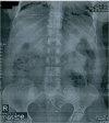
The intrauterine device (IUD) is the most common method of reversible contraception in women. However, IUD can perforate the uterus and also migrate into pelvic or abdominal organs. A 43-year-old woman with a 5-year history of IUD placement and without specific symptoms, decided to remove her IUD and undergo tubal ligation. Radiological assessment, including a pelvic X-ray and ultrasonography, revealed no copper IUD within the uterus. Retrieval attempts with cystoscopy were unsuccessful. The IUD was found embedded in the fundal part of the bladder wall and was subsequently removed through a laparotomy incision. Although there are cases in the literature that were successfully managed with cystoscopy, in chronic cases, the formation of granulation tissue may preclude retrieval of an IUD using this intervention.
Keywords: Bladder; Contraception; Intrauterine devices; Migration.
Conflict of interest statement
No potential conflict of interest relevant to this article was reported.
Figures


References
-
- Istanbulluoglu MO, Ozcimen EE, Ozturk B, Uckuyu A, Cicek T, Gonen M. Bladder perforation related to intrauterine device. J Chin Med Assoc. 2008;71:207–9. - PubMed
-
- Faculty of Family Planning and Reproductive Health Care. Royal College of Obstetricians and Gynecologists . UK selected practice recommendations for contraceptive use. London: FFPRHC and RCOG; 2003.
LinkOut - more resources
Full Text Sources

