. 2020 Jul 13;12(7):1883.
doi: 10.3390/cancers12071883.

Fast, In Vivo Model for Drug-Response Prediction in Patients with B-Cell Precursor Acute Lymphoblastic Leukemia

Affiliations

Fast, In Vivo Model for Drug-Response Prediction in Patients with B-Cell Precursor Acute Lymphoblastic Leukemia

Anton Gauert et al. Cancers (Basel). .

Abstract

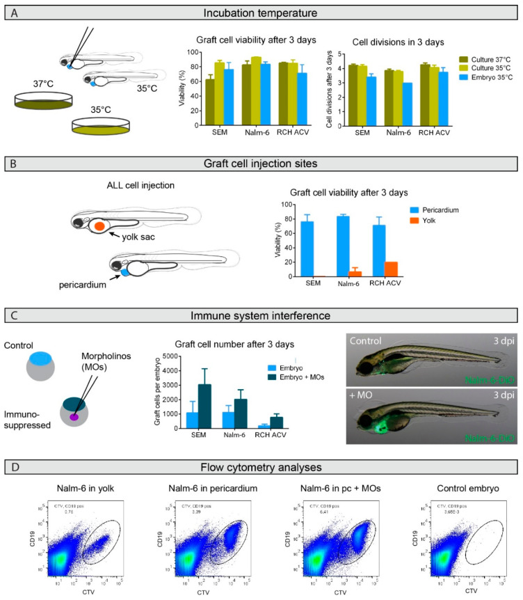
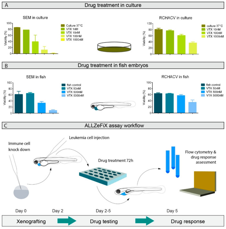
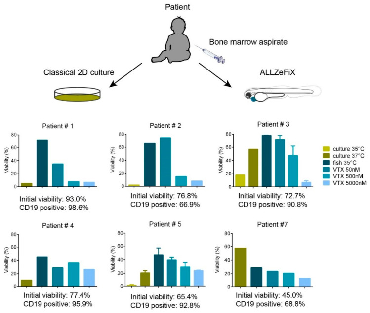
Only half of patients with relapsed B-cell precursor (BCP) acute lymphoblastic leukemia (ALL) currently survive with standard treatment protocols. Predicting individual patient responses to defined drugs prior to application would help therapy stratification and could improve survival. With the purpose to aid personalized targeted treatment approaches, we developed a human-zebrafish xenograft (ALL-ZeFiX) assay to predict drug response in a patient in 5 days. Leukemia blast cells were pericardially engrafted into transiently immunosuppressed Danio rerio embryos, and engrafted embryos treated for the test case, venetoclax, before single-cell dissolution for quantitative whole blast cell analysis. Bone marrow blasts from patients with newly diagnosed or relapsed BCP-ALL were successfully expanded in 60% of transplants in immunosuppressed zebrafish embryos. The response of BCP-ALL cell lines to venetoclax in ALL-ZeFiX assays mirrored responses in 2D cultures. Venetoclax produced varied responses in patient-derived BCP-ALL grafts, including two results mirroring treatment responses in two refractory BCP-ALL patients treated with venetoclax. Here we demonstrate proof-of-concept for our 5-day ALL-ZeFiX assay with primary patient blasts and the test case, venetoclax, which after expanded testing for further targeted drugs could support personalized treatment decisions within the clinical time window for decision-making.

Keywords: BCP-ALL; patient-derived xenograft; pediatric cancer; personalized therapy; zebrafish.


Conflict of interest statement

The authors declare no conflict of interest.

Figures

Figure 1
Figure 1
Successful B-cell precursor acute lymphoblastic leukemia (BCP-ALL) graft expansion in zebrafish embryos depends on graft site and host immunosuppression but not on temperature. (A). Viability and growth rate were flow cytometrically assessed for graft cells from groups of 10–20 zebrafish embryos at 35 °C or classical in vitro cultures at 35 °C and 37 °C from three or four independent experiments. (B). Graft cell viability was flow cytometrically assessed 3 days after injection into either the yolk sac (orange) or pericardium (blue). The two independent experiments performed for injections of Nalm-6 into the yolk sac compared to the four experiments with pericardium injections produced a significant difference, with a p-value of 0.007 in an unpaired t-test. Injections were performed in groups of 10–20 embryos. (C). Host embryos were immunosuppressed by morpholino (MO) injection into fertilized eggs (dark blue) and compared to untreated controls (light blue). Graft cell number per host embryo was calculated from flow cytometric analyses from groups of 10–20 embryos in three or four independent experiments. The comparisons of immunosuppressed injections with untreated controls produced p-values in a paired t-test: p = 0.045 for SEM cells, p = 0.095 for Nalm-6 cells and p = 0.049 for RCH-ACV cells. Bars represent means ± SEM. Microscopic images show 5-day old host embryos with (+MO) or without (control) immunosuppression 3 days post-injection (dpi) with DiO-labeled Nalm-6 cell injections into the pericardium. Only one biological replicate was performed for SEM and RCH-ACV injections into the yolk sac. Representative images shown. (D). Representative flowcytometric scatter plots of Nalm-6 cells following engraftment in zebrafish embryos. CD19 positive Nalm-6 cells prelabeled with CellTrace Violet can be separated from auto-fluorescent zebrafish cells to sort out the graft cell population for analysis. Engraftment site indicated as well as whether the host embryo was transiently immunosuppressed using morpholinos (MOs). Groups of 10 embryos from each treatment group were pooled before single-cell dissociation for flow cytometric analysis. Control embryos not engrafted show auto-fluorescence. For details see also Figure S1. Pc = pericardium.
Figure 2
Figure 2
Optimized human–zebrafish xenograft (ALL-ZeFiX) assay reveals graft response to venetoclax (VTX) comparable to results from conventional in vitro treatment. (A). Viability of SEM and RCH-ACV cells treated for 48 h with indicated concentrations of venetoclax at 37 °C from two or three independent experiments in conventional 2D culture assessed by flow cytometry. (B). Viability of SEM and RCH-ACV cell grafts in immunosuppressed embryos treated with indicated concentrations of venetoclax at 35 °C for 72 h from three independent experiments assessed by flow cytometry. Each experiment was measured as the mean of a pool of 10–20 embryos. Bars represent means ± SEM. (C). Workflow is diagrammatically shown for the ALL-ZeFiX assay.
Figure 3
Figure 3
Primary BCP-ALL patient samples can be expanded in zebrafish embryos with individual responsiveness to venetoclax. Viability of isolated mononuclear cells from primary bone marrow aspirates engrafted in immunosuppressed zebrafish embryos or in vitro 2D cultures as controls were assessed flow cytometrically after 3 days of exposure to the range of venetoclax concentrations. Graphs for patients 1, 2, 4 and 7 represent one experiment measured as the mean of a pool of 10–20 embryos. Graphs for patients 3 and 5 represent the means from two independent experiments. Error bars represent SEM.

References

    1. Hunger S.P., Mullighan C.G. Acute lymphoblastic leukemia in children. N. Engl. J. Med. 2015;373:1541–1552. doi: 10.1056/NEJMra1400972. - DOI - PubMed
    1. Lanza F., Maffini E., Rondoni M., Massari E., Faini A.C., Malavasi F. CD22 expression in b-cell acute lymphoblastic leukemia: Biological significance and implications for inotuzumab therapy in adults. Cancers. 2020;12:303. doi: 10.3390/cancers12020303. - DOI - PMC - PubMed
    1. Inaba H., Pui C.H. Immunotherapy in pediatric acute lymphoblastic leukemia. Cancer Metastasis Rev. 2019;38:595–610. doi: 10.1007/s10555-019-09834-0. - DOI - PMC - PubMed
    1. Frismantas V., Dobay M.P., Rinaldi A., Tchinda J., Dunn S.H., Kunz J., Richter-Pechanska P., Marovca B., Pail O., Jenni S., et al. Ex vivo drug response profiling detects recurrent sensitivity patterns in drug-resistant acute lymphoblastic leukemia. Blood. 2017;129:e26–e37. doi: 10.1182/blood-2016-09-738070. - DOI - PMC - PubMed
    1. Worst B.C., van Tilburg C.M., Balasubramanian G.P., Fiesel P., Witt R., Freitag A., Boudalil M., Previti C., Wolf S., Schmidt S., et al. Next-generation personalised medicine for high-risk paediatric cancer patients—The INFORM pilot study. Eur. J. Cancer. 2016;65:91–101. doi: 10.1016/j.ejca.2016.06.009. - DOI - PubMed