

Accuracy of Dynamic Navigation System Workflow for Implant Supported Full Arch Prosthesis: A Case Series
- PMID: 32668790
- PMCID: PMC7400369
- DOI: 10.3390/ijerph17145038
Accuracy of Dynamic Navigation System Workflow for Implant Supported Full Arch Prosthesis: A Case Series
Abstract
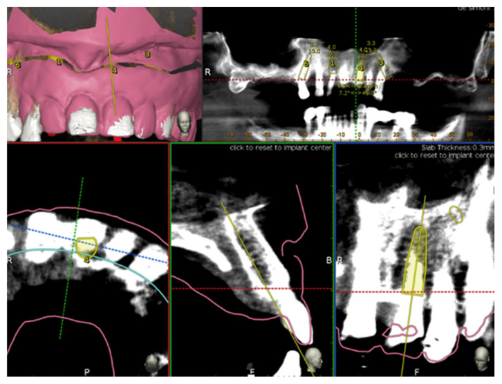
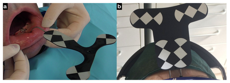
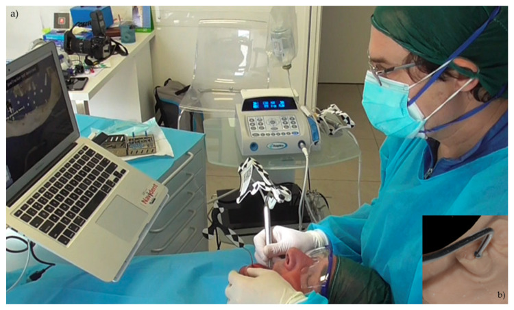
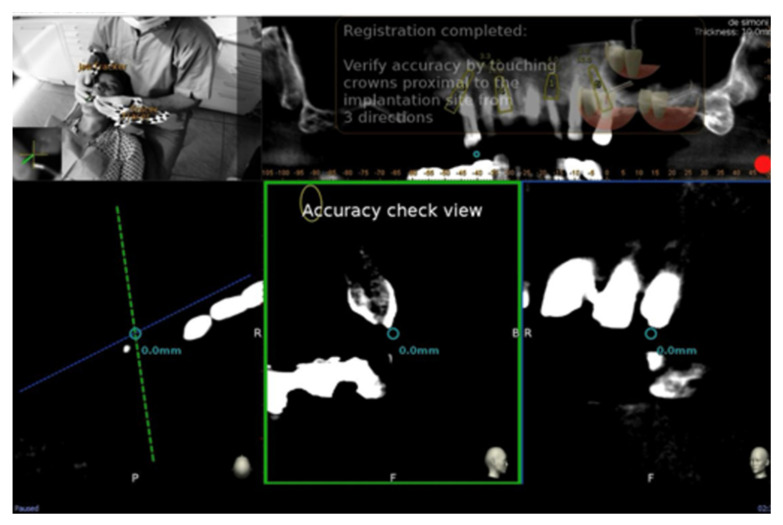
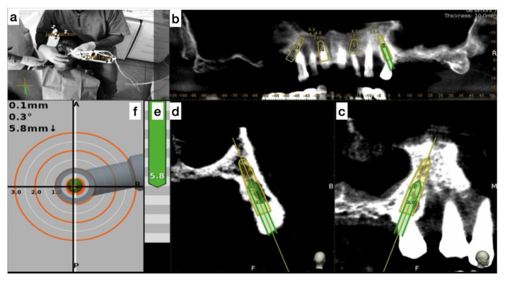
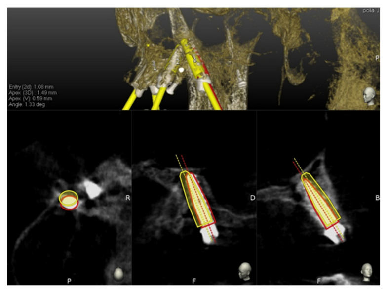
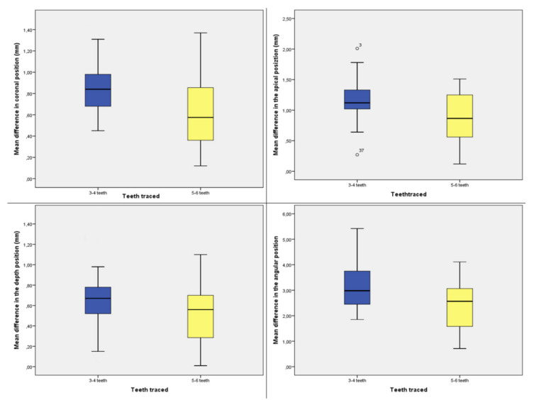
A minimally invasive implant treatment approach for future full arch implant prosthetic rehabilitations of trophic jaws represents a challenge. An optimal implant planning is strongly related with an accurate merge of the prosthetic information and the radiographic data. To comply with that, most computer aided implantology (CAI) systems require additional steps, as radiographic stents or fiducial markers to overlap digital jaw scans to cone beam computed tomography (CBCT) data. Using dynamic CAI, residual teeth (up to three) make it possible for the merge to avoid new radiographic scans. An additional challenge is the treatment involving immediate implants compared with delayed implants placed into healed bone. As for other static CAI systems, the operator's experience and the quality of the CBCT data make the planning affordable and secure the entire implants placement procedure. The literature reports accuracies in terms of comparison between placed implants and planned implants, following a double CBCT approach, based on radiographic volume overlapping. Thirteen consecutive future totally edentulous patients (77 implants), divided into two groups (group A: 3-4 teeth traced; group B: 5-6 teeth traced) requiring a full arch implant prosthetic rehabilitation were included in the reported case series. A dynamic CAI was used to plan and to place all implants following all the recommended digital steps. The software used provided a tool (Trace and Place) that made the merge between X-ray views of the residual teeth and their own positions possible. This method definitely registered that teeth positions comply with the required accuracy live check. After implants placement, a post-operative CBCT was taken in order to evaluate the deviations of the achieved implants at coronal, apical, and depth level as well as angular deviations. Statistically significant radiological mean difference between the two groups was found in the coronal position of implants (0.26 mm, p < 0.001), in the apical position of implants (0.29 mm, p < 0.001), in the depth of implants (0.16 mm, p = 0.022), and in the angular deviation (0.7, p = 0.004). The use of the TaP technology for the treatment of the patients with at least three stable teeth that need to be removed for a totally implant prosthetic treatment is a promising technique. The performed accuracy analysis demonstrated that this digital protocol can be used without a loss of accuracy of the achieved implants compared to planned ones.
Keywords: atrophic maxilla; computer aided implantology; navigation implantology; totally edentulous patients.
Conflict of interest statement
The authors declare no conflict of interest.
Figures

References
-
- La Monaca G., Iezzi G., Cristalli M.P., Pranno N., Sfasciotti G.L., Vozza I. Comparative Histological and Histomorphometric Results of Six Biomaterials Used in Two-Stage Maxillary Sinus Augmentation Model after 6-Month Healing. Biomed. Res. Int. 2018;2018:9430989. doi: 10.1155/2018/9430989. - DOI - PMC - PubMed
-
- Borges T.F., Mendes F.A., De Oliveira T.R., Gomes V.L., do Prado C.J., das Neves F.D. Mandibular overdentures with immediate loading: Satisfaction and quality of life. Int. J. Prosthodont. 2011;24:534–539. - PubMed
Publication types
MeSH terms
LinkOut - more resources
Full Text Sources
Research Materials
Miscellaneous
