. 2020 Nov 1;26(21):5759-5771.
doi: 10.1158/1078-0432.CCR-20-0251. Epub 2020 Jul 15.

Human Cancers Express TRAILshort, a Dominant Negative TRAIL Splice Variant, Which Impairs Immune Effector Cell Killing of Tumor Cells

Affiliations

Human Cancers Express TRAILshort, a Dominant Negative TRAIL Splice Variant, Which Impairs Immune Effector Cell Killing of Tumor Cells

Fatma Aboulnasr et al. Clin Cancer Res. .

Abstract

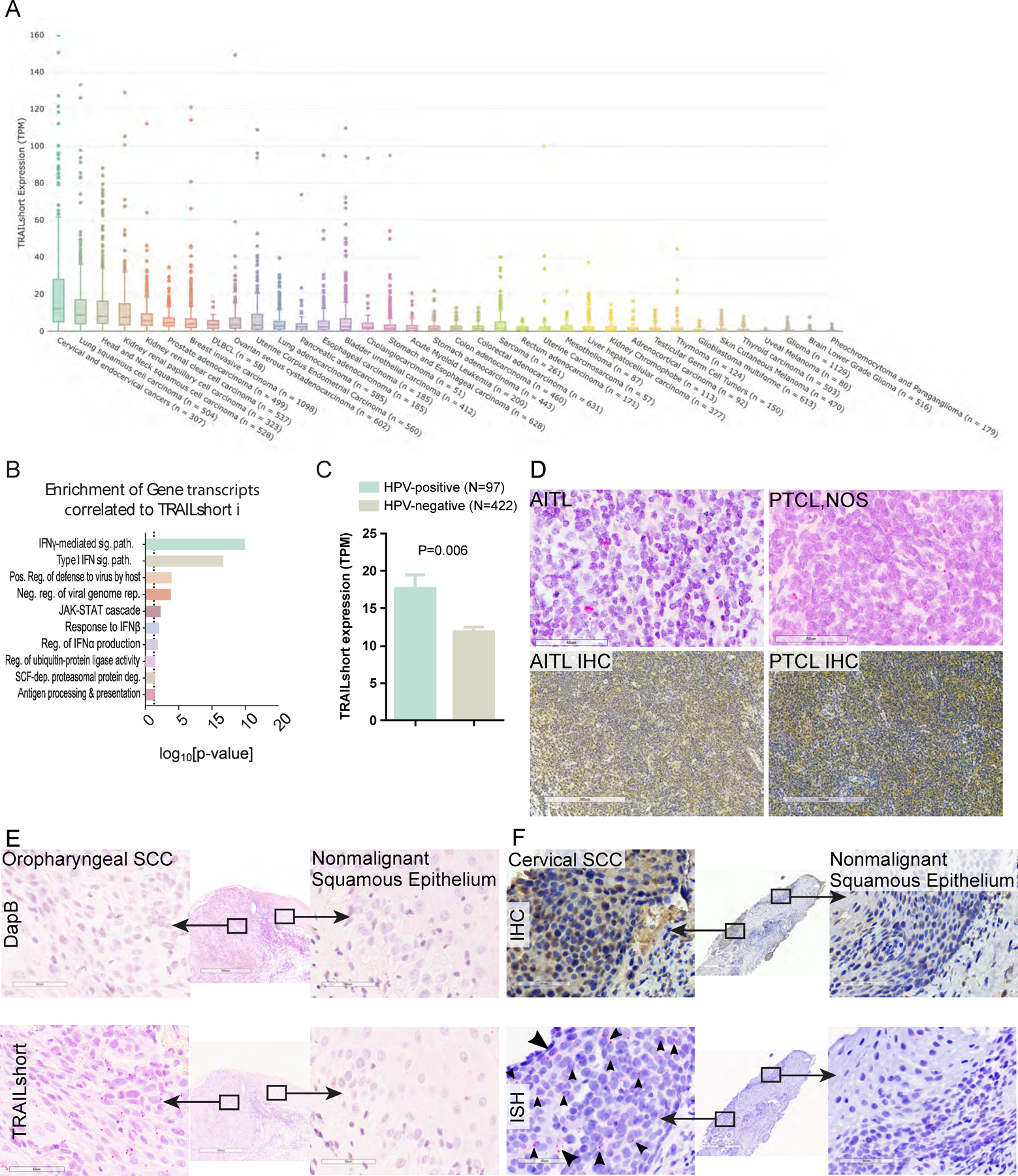
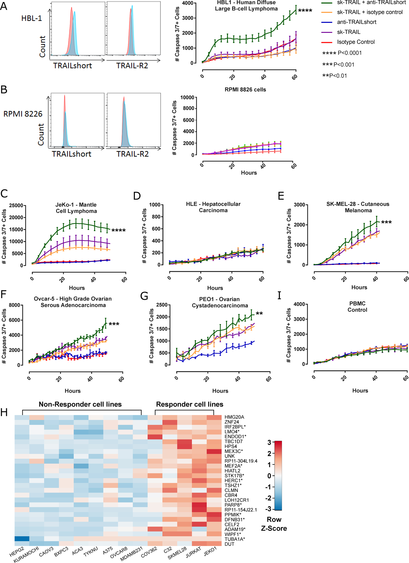
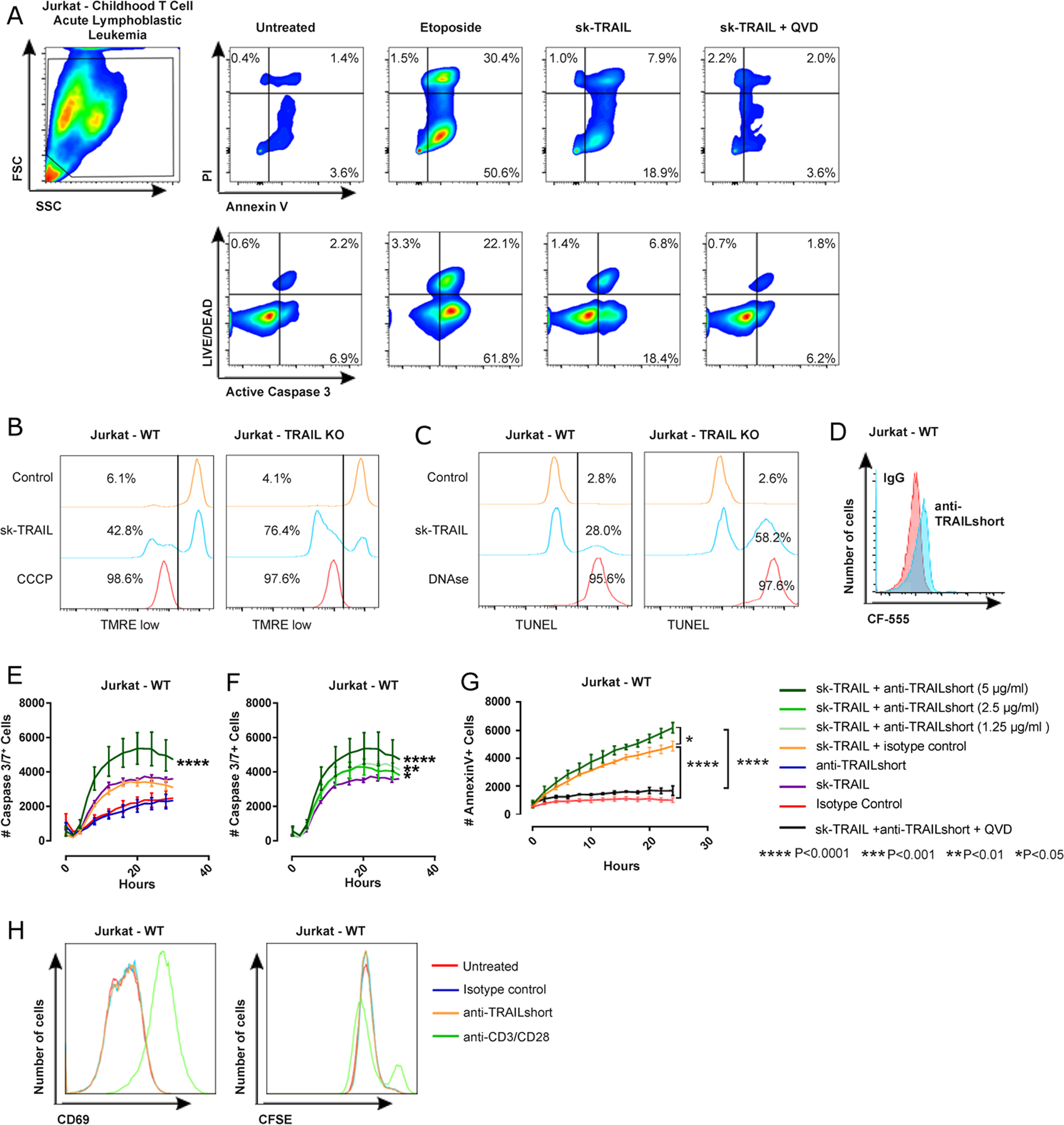
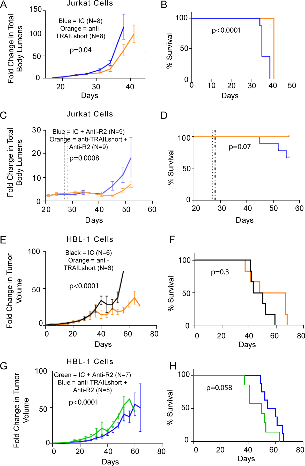
Purpose: TNF-related apoptosis inducing ligand (TRAIL) expression by immune cells contributes to antitumor immunity. A naturally occurring splice variant of TRAIL, called TRAILshort, antagonizes TRAIL-dependent cell killing. It is unknown whether tumor cells express TRAILshort and if it impacts antitumor immunity.

Experimental design: We used an unbiased informatics approach to identify TRAILshort expression in primary human cancers, and validated those results with IHC and ISH. TRAILshort-specific mAbs were used to determine the effect of TRAILshort on tumor cell sensitivity to TRAIL, and to immune effector cell dependent killing of autologous primary tumors.

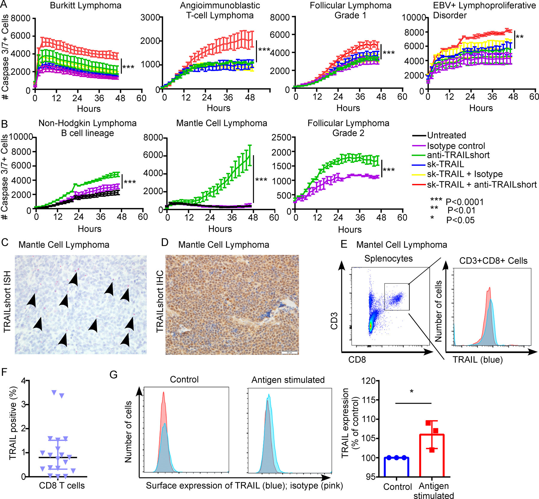
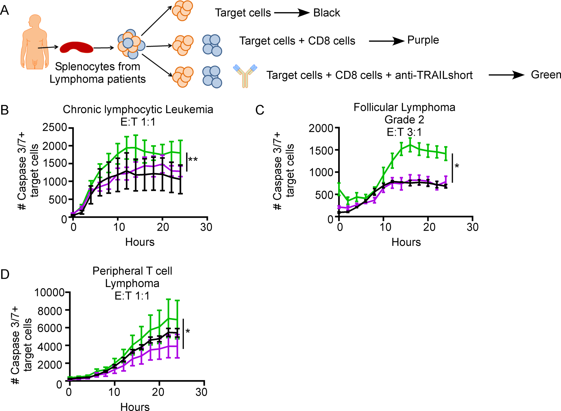
Results: As many as 40% of primary human tumors express TRAILshort by both RNA sequencing and IHC analysis. By ISH, TRAILshort expression is present in tumor cells and not bystander cells. TRAILshort inhibition enhances cancer cell lines sensitivity to TRAIL-dependent killing both in vitro and in immunodeficient xenograft mouse models. Immune effector cells isolated from patients with B-cell malignancies killed more autologous tumor cells in the presence compared with the absence of TRAILshort antibody (P < 0.05).

Conclusions: These results identify TRAILshort in primary human malignancies, and suggest that TRAILshort blockade can augment the effector function of autologous immune effector cells.See related commentary by de Miguel and Pardo, p. 5546.


Conflict of interest statement

Conflict of Interest:

One or more of the investigators associated with this project and Mayo Clinic have a Financial Conflict of Interest in technology used in the research and that the investigator(s) and Mayo Clinic may stand to gain financially from the successful outcome of the research and this research has been reviewed by the Mayo Clinic Conflict of Interest Review Board and is being conducted in compliance with Mayo Clinic Conflict of Interest policies.

Disclosures unrelated to this work: SSK is inventor on patents in CART cell therapy that are licensed to Novartis (through an agreement between Mayo Clinic, University of Pennsylvania, and Novartis), Humanigen (through Mayo Clinic), and Mettaforge (through Mayo Clinic). SSK receives research funding from Kite, Gilead, Novartis, Juno, Celgene, Morphosys, Humanigen, Tolero, and Lentigen. ADB is a consultant and receives consulting fees or equity shares from Abbvie, Nference, and Xentalis. ADB is a founder and president of Splissen Therapeutics which has licensed patents related to TRAILshort.

Figures

Figure 1.
Figure 1.. TRAILshort is prevalent within human cancer tissues.
(A) Tissues within the TCGA dataset were queried for the presence of TRAILshort transcripts, and data are expressed as transcripts per million (TPM) according to tissue type. (B) Pearson’s correlation coefficients (PCC) identified 66 gene transcripts co-associated with expression of TRAILshort transcripts, and the functional clustering is shown. (C) TRAILshort transcripts were compared between HPV-positive (N=97) and HPV-negative (N=422) tumors in the TCGA dataset; depicted are mean +/− standard error; P=0.0061 by two-tailed Mann-Whitney test. (D-F) The indicated tissues were stained by immunohistochemistry (IHC) for TRAILshort protein expression (brown staining), or by in situ hybridization (ISH) for TRAILshort mRNA expression (red dots indicated with black arrowheads). (D) Angioimmunoblastic T-cell lymphoma (AITL) and peripheral T-cell lymphoma not otherwise specified (PTCL NOS) stain positive for TRAILshort mRNA ( red dots with black arrowheads). (top panels, RNA ISH, scale bar = 60 µm) as well as by IHC (bottom panels, scale bar = 300 µm). (E) Oropharyngeal squamous cell carcinoma (SCC) tissue was stained by ISH using a negative control probe, DapB top panels, and no signal is detected, yet TRAILshort mRNA is detected by ISH in the malignant tissue indicated by the red dots (highlighted by black arrowheads), but not in the non-malignant adjacent tissue Scale bar = 60 µm for all panels. (F) TRAILshort protein detected by IHC (top panels) or TRAILshort mRNA detected by ISH (bottom panels) in malignant (left panels) or non-malignant (right panels) cervical squamous epithelium.
Figure 2.
Figure 2.. Neutralization of TRAILshort enhances TRAIL induced cell killing
Human cancer cell lines were assessed for TRAILshort and TRAIL receptor 2 expression, and in parallel tested for cell killing in response to skTRAIL, with or without TRAILshort antibody. TRAILshort and TRAIL receptor 2 expression is shown for (A) HBL1 cells or (B) RPMI 8226 cells (Red histogram= isotype control staining; blue =TRAILshort or TRAILR2), and these cells were tested for cell killing in response to recombinant TRAIL (skTRAIL) alone, anti TRAILshort antibody alone, or the combination. Cell killing curves performed as in A and B, shown for (C) JeKo-1, (D) HLE, (E) SK-MEL-28, (F) Ovcar-5, and (G) PEO1, (I) primary peripheral blood mononuclear cells (PBMCs). (H) The transcriptional profile of anti-TRAILshort-responsive cell lines was compared to that of anti-TRAILshort-non-responsive cell lines to generate a heat map, which identified differentially expressed genes. Interferon response genes are indicated by an asterisk.
Figure 3.
Figure 3.. Neutralization of TRAILshort does not alter cell proliferation nor activation, but enhances apoptotic cell death.
(A) Jurkat T cells were treated with control, sk-TRAIL, or sk-TRAIL plus the pan-caspase inhibitor QVD for 24 hours and assessed for apoptosis by Annexin V/PI staining and active caspase 3/ LIVE/DEAD staining by flow cytometry. Etoposide was used as a positive control. (B) Wild type (WT) Jurkat T cells or Jurkat T cells genetically deficient in TRAIL by CRISPR (TRAIL KO) were treated with skTRAIL or vehicle control for 12 hours and mitochondrial depolarization assessed by tetramethylrhodamine, ethyl ester, perchlorate (TMRE) staining and flow cytometry. Carbonyl cyanide m-chlorophenylhydrazone (CCCP) was used as a positive control. Percentage of TMRE low cells is enumerated. (C) WT and TRAIL KO Jurkat T cells were treated with skTRAIL or control for 12 hours and DNA fragmentation assessed by TUNEL staining and flow cytometry. DNAse treatment was used as positive control. The percentage of TUNEL positive cells is enumerated. (D) Jurkat T cells were assessed for surface expression of TRAILshort by flow cytometry. (E – F) Ten thousand Jurkat T cells were treated with either an isotype control antibody (5µg/ml), recombinant pre-aggregated sk-TRAIL (1 ng/ml, unless unresponsive in which case 10 ng/ml was used), mouse anti-TRAILshort antibody (5 µg/ml, unless otherwise noted), or a combination of isotype control and either sk-TRAIL or mouse anti-TRAILshort antibody at low, medium, or high concentration (1.25, 2.5 or 5 µg/ml), plus sk-TRAIL as indicated. The number of caspase 3/7 positive cells was monitored every 2 hours. All experiments were performed in triplicate. Data are represented as mean ± SE. Significance shown in A and B compares sk-TRAIL + anti-TRAILshort versus sk-TRAIL alone. (G) Jurkat cells were treated with sk-TRAIL plus anti-TRAILshort antibody in the presence of or absence of the pan-caspase inhibitor Q-VD to test the caspase-dependence of apoptosis. (H) WT Jurkat cells were treated with nothing, isotype control, anti-TRAILshort antibody or anti-CD3/CD28 microbeads and assessed for surface expression of CD69 (at 3 days) and CFSE dilution (at 5 days).
Figure 4.
Figure 4.. Anti-TRAILshort antibody alone or in combination with a TRAIL agonist has antitumor effects in Jurkat T cell and HBL-1 xenograft models.
(A-B) Non-obese diabetic, severe combined-immunodeficient, common γ-chain deficient (NSG) mice were implanted with Jurkat human T leukemia cells expressing luciferase by IV injection, and tumors were allowed to become established. Mice were treated weekly with mouse anti-TRAILshort antibody (clone 2.2) alone or isotype control antibody. Mice were imaged twice weekly and followed for (A) total body luminescence over time and (B) survival. (C-D) Mice injected with luciferase expressing Jurkat T cells were administered a single injection of mouse anti-TRAILshort antibody (clone 2.2) followed 24 hours later by a single injection of anti-TRAIL-R2 antibody, and followed for (C) mouse whole body luminescence over time (P=0.0008) and (D) Mouse survival (P=0.07). (E-H) NSG mice were implanted with HBL-1 cells by IV injection, and tumors were allowed to become established. These mice were treated weekly with anti-TRAIL short antibody or isotype control alone (E-F), or followed the next day by anti-TRAILR2 antibody (G-H), and tumors measured and followed for (E and G) tumor size and (F and H) survival. Results shown are mean ± SD.
Figure 5.
Figure 5.. Humanized anti-TRAILshort antibodies have antitumor activity in primary human tumor samples ex vivo.
Ten thousand splenocytes harvested from human patients with suspected malignancies were treated within 4 hours of harvest with isotype control antibody (5µg/ml), recombinant pre-aggregated sk-TRAIL (1 ng/ml), humanized anti-TRAILshort antibody (5 µg/ml), the combination of isotype control antibody and sk-TRAIL, or humanized anti-TRAILshort antibody at low medium or high dose (1.25, 2.5 or 5 µg/ml) plus sk-TRAIL as indicated. (A) Representative patient samples in which splenocyte death was augmented by the addition of anti-TRAILshort antibody to the TRAIL agonist skTRAIL. (B) Representative patient samples in which splenocyte death was induced with anti-TRAILshort antibody treatment alone. (C) Splenic tissue from the patient with Mantle cell lymphoma from B, was stained for TRAILshort expression by ISH (red dots and arrows), and (D) IHC ( brown staining). (E) CD8 T cells from the splenocyte suspension were analyzed by flow cytometry using CD3/CD8 and TRAIL antibodies; TRAIL expression in CD3+CD8+ cells is shown. (F) Surface TRAIL expression was examined in peripheral CD3+CD8+ T cells from healthy donors by flow cytometry, in unstimulated conditions or (G) stimulated with tetanus toxoid (recall antigen) ex vivo for 24 hours, histogram shows pooled results from 3 donors.
Figure 6.
Figure 6.. Humanized anti-TRAILshort antibodies augment antitumor activity of immune effector cells ex vivo.
(A) CD8 positive cells were positively selected from bulk patient splenocytes, and co-cultured with autologous target cells (bulk splenocytes after CD8 cell removal stained with cell tracker red) at the indicated effector to target ratios, in the presence or absence of anti-TRAILshort antibody, and cell death was measured over time in the red target cell population. (B-D) Representative data of the number of Caspase 3/7 positive target cells in co-culture from three patient samples with response to anti-TRAILshort antibody.

Comment in

References

    1. Walczak H, et al., Tumoricidal activity of tumor necrosis factor-related apoptosis-inducing ligand in vivo. Nat Med, 1999. 5(2): p. 157–63. - PubMed
    1. Mirandola P, et al., Activated human NK and CD8+ T cells express both TNF-related apoptosis-inducing ligand (TRAIL) and TRAIL receptors but are resistant to TRAIL-mediated cytotoxicity. Blood, 2004. 104(8): p. 2418–24. - PubMed
    1. Sedger LM, et al., Characterization of the in vivo function of TNF-alpha-related apoptosis-inducing ligand, TRAIL/Apo2L, using TRAIL/Apo2L gene-deficient mice. Eur J Immunol, 2002. 32(8): p. 2246–54. - PubMed
    1. Zerafa N, et al., Cutting edge: TRAIL deficiency accelerates hematological malignancies. J Immunol, 2005. 175(9): p. 5586–90. - PubMed
    1. Cretney E, et al., Increased susceptibility to tumor initiation and metastasis in TNF-related apoptosis-inducing ligand-deficient mice. J Immunol, 2002. 168(3): p. 1356–61. - PubMed

Publication types

Substances