. 2020 Oct;61(10):1308-1319.
doi: 10.1194/jlr.RA119000479. Epub 2020 Jul 15.

Interleukin 6 reduces allopregnanolone synthesis in the brain and contributes to age-related cognitive decline in mice

Affiliations

Interleukin 6 reduces allopregnanolone synthesis in the brain and contributes to age-related cognitive decline in mice

Eileen E Parks et al. J Lipid Res. 2020 Oct.

Abstract

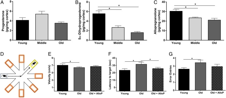
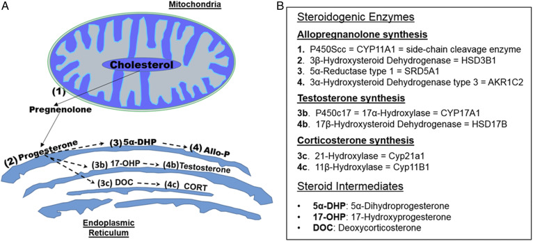
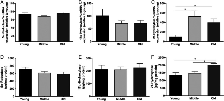
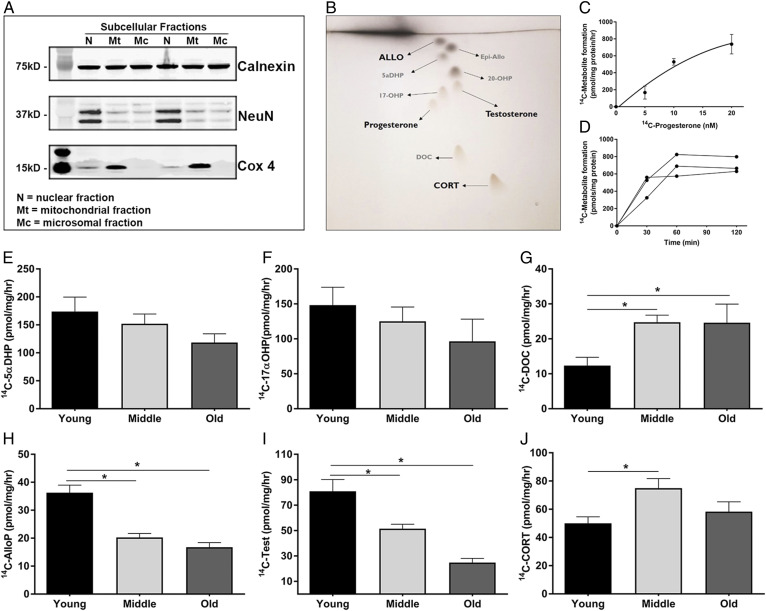
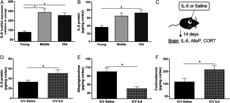
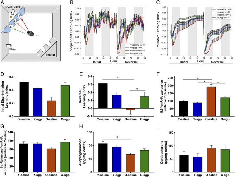
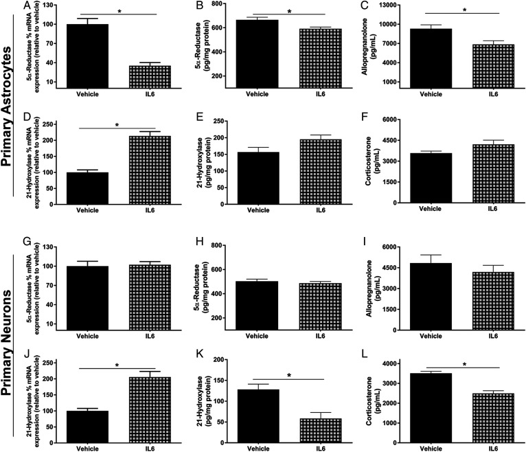
Cognitive decline with age is a harmful process that can reduce quality of life. Multiple factors have been established to contribute to cognitive decline, but the overall etiology remains unknown. Here, we hypothesized that cognitive dysfunction is mediated, in part, by increased levels of inflammatory cytokines that alter allopregnanolone (AlloP) levels, an important neurosteroid in the brain. We assessed the levels and regulation of AlloP and the effects of AlloP supplementation on cognitive function in 4-month-old and 24-month-old male C57BL/6 mice. With age, the expression of enzymes involved in the AlloP synthetic pathway was decreased and corticosterone (CORT) synthesis increased. Supplementation of AlloP improved cognitive function. Interestingly, interleukin 6 (IL-6) infusion in young animals significantly reduced the production of AlloP compared with controls. It is notable that inhibition of IL-6 with its natural inhibitor, soluble membrane glycoprotein 130, significantly improved spatial memory in aged mice. These findings were supported by in vitro experiments in primary murine astrocyte cultures, indicating that IL-6 decreases production of AlloP and increases CORT levels. Our results indicate that age-related increases in IL-6 levels reduce progesterone substrate availability, resulting in a decline in AlloP levels and an increase in CORT. Furthermore, our results indicate that AlloP is a critical link between inflammatory cytokines and the age-related decline in cognitive function.

Keywords: Alzheimer’s disease; aging; cognitive function; enzyme regulation; inflammation; neurosteroid; progesterone; steroid hormones.


Conflict of interest statement

Conflict of interest—The authors declare that they have no conflicts of interest with the contents of this article.

Figures

Fig. 1.
Fig. 1.
AlloP decreases in the brain with age, and its restoration improves working memory. A–C: LC/MS quantification of PRG (A), 5α-dihydroprogesterone (B), and AlloP (C) in the cerebral cortex from young (3 months), middle-aged (12 months), and old mice (24 months) (n = 6 per group; one-way ANOVA). D: Illustration of the RAWM. Orange boxes indicate incorrect arms, and the yellow box indicates the target arm containing the escape platform. E–G: Performance in the RAWM in young (3 months), old (24 months), and old mice treated with AlloP. E: Average velocity of the mice in each group while in the RAWM. F: Average latency from the starting arm to the target platform. G: Average entries into incorrect arms. E–G: Data represent the average of eight trials per day over 3 days (n = 9 per group; two-way ANOVA and Tukey HSD pairwise comparisons). *P < 0.05. Statistics were adjusted for multiple comparisons. Only significant differences are depicted in the figure. Data are presented as mean ± SEM.
Fig. 2.
Fig. 2.
Biosynthetic pathways leading to neurosteroid synthesis. A: Illustration of steroidogenic enzymes produced in the mitochondria and endoplasmic reticulum leading to the synthesis of AlloP, testosterone, and CORT. B: Table outlining the steroidogenic enzymes associated with the respective numbers in A. Cholesterol is the precursor for all neurosteroid synthesis. Cholesterol is first metabolized by P450Scc (1) housed in the mitochondria to form pregnenolone. After exiting the mitochondria, pregnenolone can then be metabolized by 3β-hydroxysteroid dehydrogenase (2), located in the endoplasmic reticulum, to form PRG. PRG can then be metabolized by several different enzymes to form unique steroid metabolites. AlloP is formed via 5α-reductase (3) and 3α-hydroxysteroid dehydrogenase (4), respectively. Testosterone can be formed via P450c17 (3b) and 17β-hydroxysteroid dehydrogenase (4b), respectively. CORT can also be formed from 21-hydroxylase (3c) and 11β-hydroxylase (4c), respectively.
Fig. 3.
Fig. 3.
The mRNA expression and protein levels of steroidogenic enzymes are altered with age. A–C: Percent mRNA expression of 5α-reductase, 17α-hydroxysteroid dehydrogenase, and 21-hydroylase in the cortex of young (3 months), middle-aged (12 months), and old (24 months) mice (n = 8 per group; one-way ANOVA). D–F: Protein levels (picograms per milligram of protein) of 5α-reductase, 17α-hydroxysteroid dehydrogenase, and 21-hydroylase in the cortex of young (3 months), middle-aged (12 months), and old (24 months) mice (n = 8 per group; one-way ANOVA). Statistics were adjusted for multiple comparisons. Only significant differences are depicted in the figure. Data are presented as mean ± SEM
Fig. 4.
Fig. 4.
PRG metabolism via steroidogenic enzymes are altered with age in the cerebral cortex. A: Representative Western blot showing the separation of subcellular fractions. Calnexin is a marker for the microsomal fraction, Cox4 marks the mitochondrial fraction, and NeuN marks the nuclear fraction (n = 3, with three replicate experiments). B: Representative image of the TLC plates used to separate the individual steroid metabolites. C: Dose response curve of increasing amounts 14C-PRG to 14C-metabolites in microsomes (n = 3). Data represent the average total 14C-metabolite picomoles formed per milligram of protein per hour. D: Time course of 14C-metabolite formation in microsomes following the addition of 14C-PRG (n = 3). E–J: Metabolite formation of 5α-dihydroprogesterone (5αDHP) (E), 17α-hydroxyprogesterone (17αOHP) (F), deoxycorticosterone (DOC) (G), AlloP (H), testosterone (Test) (I), and CORT (J) in microsomes isolated from the cerebral cortex of young (3 months), middle-aged (12 months), and old (24 months) mice. PRG metabolite formation is shown as the picomoles of each 14C-metabolite formed per milligram of microsomal protein per hour (n = 4 per group; one-way ANOVA) (*P < 0.05). Statistics were adjusted for multiple comparisons. Only significant differences are depicted in the figure. Data are presented as mean ± SEM
Fig. 5.
Fig. 5.
IL-6 increases in the brain with age, and its administration to young mice reduces AlloP synthesis and increases CORT synthesis. A, B: Cerebral cortex tissues from young (3 months), middle-aged (12 months), and old (24 months) mice. A: Percent mRNA expression of IL-6 across age relative to young (n = 8 per group; one-way ANOVA). B: Levels of IL-6 (picograms per milligram of protein) in the cortex across age (n = 6 per group; one-way ANOVA). C: Experimental design of saline or IL-6 infusions to the mouse brain. Saline or IL-6 was loaded into an osmotic pump that was coupled to a cannula and inserted into the lateral ventricle of the brain in young (4 months) mice. The osmotic pump delivered saline or IL-6 (100 ng/day) continuously for 14 days. D: Levels of IL-6 (pg/mg protein) in the cortex following Saline or IL-6 intracerebroventricular infusion. E, F: AlloP and CORT levels in the cortex of young mice following 2 weeks of intracerebroventricular infusions of either saline or IL-6 (100 ng/day) to the lateral ventricle of the brain (n = 8 per group, unpaired Student’s t-test, *P < 0.05). Statistics were adjusted for multiple comparisons. Only significant differences are depicted in the figure. Data are presented as mean ± SEM.
Fig. 6.
Fig. 6.
The effects of IL-6 inhibition on neurosteroid synthesis and working memory. A: Illustration of the PhenoTyper home cage system containing the cognition wall used to assess working memory. On days 1–2, the initial learning period, the mouse must learn to enter the left most entry of the cognition wall at least five times to receive a food pellet. On days 3–4, the reversal period, the mouse learns to enter the right most entry at least five times to receive a food pellet. B, C: Working memory assessment in young and old mice following a 2 week intracerebroventricular infusion of either saline or sgp130 (IL-6 inhibitor) to the lateral ventricle of the brain. Learning indices are calculated by (correct entries – incorrect entries)/total entries made per hour. B: The independent index represents the average learning index per group across each hour of the initial learning and reversal periods. C: The cumulative index represents the average learning index per group accumulated over consecutive hours in both the initial learning period and the reversal period. D: Average learning indices per group during the initial discrimination period. E: Average learning indices per group during the reversal period (n = 5–8 per group, two-way ANOVA, Tukey HSD pairwise comparisons, *P < 0.05). F–I: Cerebral cortex tissues from young and old mice treated with saline or the IL-6 inhibitor, sgp130 (n = 5–8 per group, two-way ANOVA, *P < 0.05). F: Percent mRNA expression of IL-6 across groups relative to young. G: Percent mRNA expression of 5α-reductase across groups relative to young. H, I: AlloP and CORT levels in the cortex following saline or sgp130 treatment. Statistics were adjusted for multiple comparisons. Only significant differences are depicted in the figure. Data are presented as mean ± SEM.
Fig. 7.
Fig. 7.
IL-6 reduces the synthesis of AlloP specifically in astrocytes and reduces the synthesis of CORT specifically in neurons. Primary astrocyte and neuronal cultures were treated with or without IL-6 (100 ng/ml) for 24 h and used for the following experiments. Cell culture lysates were used for percent mRNA expression (relative to vehicle control) and protein levels of 5α-reductase and 21-hydroxylase. Cell culture medium was used for AlloP and CORT quantification. 5α-Reductase mRNA expression in astrocytes and neurons (A, G), 5α-reductase protein levels in astrocytes and neurons (B, H), AlloP in cell culture media from astrocytes and neurons (C, I), 21-hydroxylase mRNA expression in astrocytes and neurons (D, J), 21-hydroxylase protein levels in astrocytes and neurons (E, K), CORT in cell culture media from astrocytes and neurons (F, L) (n = 4 replicate experiments; Student’s unpaired t-test; Bonferroni method for multiple comparisons, *P < 0.05). Data are presented as mean ± SEM.

References

    1. Cansino S., Torres-Trejo F., Estrada-Manilla C., Martinez-Galindo J. G., Hernandez-Ramos E., Ayala-Hernandez M., Gomez-Fernandez T., Ramirez-Gonzalez M. D., and Ruiz-Velasco S.. 2018. Factors that positively or negatively mediate the effects of age on working memory across the adult life span. Geroscience. 40: 293–303. - PMC - PubMed
    1. Godbout J. P., and Johnson R. W.. 2009. Age and neuroinflammation: a lifetime of psychoneuroimmune consequences. Immunol. Allergy Clin. North Am. 29: 321–337. - PubMed
    1. Chinta S. J., Woods G., Rane A., Demaria M., Campisi J., and Andersen J. K.. 2015. Cellular senescence and the aging brain. Exp. Gerontol. 68: 3–7. - PMC - PubMed
    1. Di Benedetto S., Muller L., Wenger E., Duzel S., and Pawelec G.. 2017. Contribution of neuroinflammation and immunity to brain aging and the mitigating effects of physical and cognitive interventions. Neurosci. Biobehav. Rev. 75: 114–128. - PubMed
    1. Compagnone N. A., and Mellon S. H.. 2000. Neurosteroids: biosynthesis and function of these novel neuromodulators. Front. Neuroendocrinol. 21: 1–56. - PubMed