. 2020 Aug;19(8):553-571.
doi: 10.1038/s41573-020-0071-y. Epub 2020 Jul 15.

Receptor-interacting protein kinase 1 (RIPK1) as a therapeutic target

Affiliations

Receptor-interacting protein kinase 1 (RIPK1) as a therapeutic target

Lauren Mifflin et al. Nat Rev Drug Discov. 2020 Aug.

Abstract

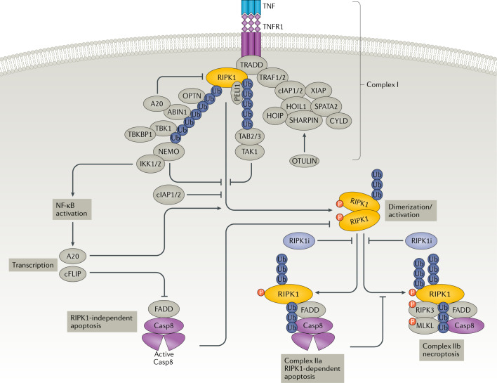
Receptor-interacting serine/threonine-protein kinase 1 (RIPK1) is a key mediator of cell death and inflammation. The unique hydrophobic pocket in the allosteric regulatory domain of RIPK1 has enabled the development of highly selective small-molecule inhibitors of its kinase activity, which have demonstrated safety in preclinical models and clinical trials. Potential applications of these RIPK1 inhibitors for the treatment of monogenic and polygenic autoimmune, inflammatory, neurodegenerative, ischaemic and acute conditions, such as sepsis, are emerging. This article reviews RIPK1 biology and disease-associated mutations in RIPK1 signalling pathways, highlighting clinical trials of RIPK1 inhibitors and potential strategies to mitigate development challenges.


Conflict of interest statement

J.Y. is a consultant for Denali Therapeutics and Sanofi. D.O. is an employee of Sanofi.

Figures

Fig. 1
Fig. 1. RIPK1 in TNFR1 signalling.
Complex I: stimulation of TNFR1 by TNF promotes the formation of an intracellular signalling complex associated with the death domain of trimerized TNFR1 that recruits two death domain-containing proteins: adaptor protein TRADD and receptor-interacting serine/threonine-protein kinase 1 (RIPK1). TRADD recruits E3 ubiquitin ligases cIAP1/2 and XIAP to perform K63 ubiquitylation of complex I, including RIPK1 K377, which in turn recruits the LUBAC complex, comprised of HOIP, HOIL1 and SHARPIN. LUBAC mediates linear (M1) ubiquitylation of RIPK1. Deubiquitinase CYLD and its adaptor protein SPATA2 modulate M1/K63 ubiquitylation of RIPK1. M1 deubiquitinase OTULIN activates LUBAC. K63 ubiquitin chains on RIPK1 recruit TAB2/3 and TAK1. M1 ubiquitin chains on RIPK1 recruit the NEMO–IκB kinase (IKK) complex, TBK1, A20, ABIN1 (A20 binding inhibitor of NF-κB-1) and OPTN. A20 in complex I suppresses the activation of RIPK1 kinase. NF-κB activation: activation of TAK1 and the IKKs promote NF-κB pathway activation to mediate transcription of both pro-inflammatory and pro-survival genes, including A20, which modulates the ubiquitylation of RIPK1 to control its activation, and cellular FLICE-like inhibitory protein (cFLIP), which modulates activation of caspase 8 (Casp8). RIPK1-dependent apoptosis (RDA): activation of TNFR1 under A20, ABIN1, cIAP1/2, NEMO, TBK1, IKK or TAK1-deficient conditions leads to RIPK1 kinase activation. Activated RIPK1 binds to FADD and Casp8 to form complex IIa and promote activation of caspases and apoptosis. Increased levels of A20 promote the activation of RIPK1 in complex IIa. Necroptosis: inhibition of Casp8-mediated cleavage of RIPK1 promotes RIPK1 dimerization via the C-terminal death domain, which leads to its activation and the subsequent formation of the necrosome (complex IIb) comprised of RIPK1, FADD, Casp8, RIPK3 and mixed-lineage kinase domain-like pseudokinase (MLKL), which in turn executes necroptosis. RIPK1-independent apoptosis: when the NF-κB pathway is inhibited, TNF stimulation can promote the formation of a cytosolic complex with FADD and Casp8 to mediate apoptosis independent of RIPK1. P, phosphate; RIPK1i, RIPK1 inhibitor; Ub, ubiquitin. Adapted from ref., Springer Nature Limited.
Fig. 2
Fig. 2. Mechanisms of RIPK1 in neurodegenerative diseases.
Activation of receptor-interacting serine/threonine-protein kinase 1 (RIPK1) in neurodegenerative diseases may promote the death of neurons and oligodendrocytes cell-autonomously, and inflammation in microglia and astrocytes that acts non-cell-autonomously to promote neurodegeneration. Neurons and oligodendrocytes: amyotrophic lateral sclerosis (ALS)/frontotemporal dementia (FTD)-associated loss-of-function (LoF) mutations in TBK1 and OPTN and ageing-related reduction in TAK1 can promote RIPK1-dependent apoptosis and necroptosis, which can be inhibited by RIPK1 inhibitors. Parkinson disease (PD)-associated mutations in OPA1 and DRP1, ALS-associated mutations in SOD1 and the chemical stressor MPTP lead to mitochondrial dysfunction and the production of reactive oxygen species (ROS). Cytosolic ROS can modulate cysteine residues on RIPK1, which promotes activation. Microglia and astrocytes: activation of RIPK1 leads to the production of pro-inflammatory cytokines in microglia. Activation of RIPK1 in Alzheimer disease (AD) leads to increased production of CST7 and CH25H, both of which are associated with the disease-associated microglia phenotype. PD/lysosomal storage disorder (LSD)-associated genes, such as GBA and NPC1, also result in lysosomal dysfunction, which can promote RIPK1 activation, likely by promoting the accumulation of RIPK1. ALS-associated mutations in SOD1 are also known to promote mitochondrial dysfunction and ROS production, which may promote glial activation of RIPK1 in disease. MLKL, mixed-lineage kinase domain-like pseudokinase; P, phosphate; RDA, RIPK1-dependent apoptosis; RIPK1i, RIPK1 inhibitor.
Fig. 3
Fig. 3. RIPK1 contains a hydrophobic pocket amenable to small-molecule inhibition.
Receptor-interacting serine/threonine-protein kinase 1 (RIPK1) contains a unique hydrophobic pocket located between the N terminus and C terminus of the kinase domain, which allosterically regulates kinase activation. All RIPK1 inhibitors discovered to date, such as necrostatin-1s (Nec-1s) shown here, bind to this pocket and stabilize RIPK1 in an inactive conformation (PDB: 4ITH). This pocket is created owing to the outward movement of the αC-helix, resulting in the loss of an ionic pair between catalytic Lys45 and Glu63 of the αC-helix. The other side of the pocket is formed by the DLG motif in the inactive DLG-in conformation (catalytic Asp146 facing away from the active centre) and the activation segment, which immediately follows the DLG motif. Ser161 residue in the activation segment forms a critical hydrogen bond with the indole of Nec-1s. GSK′772 and other benzoxazepinones also extend into the ATP binding pocket, which may contribute to the increased affinity. Adapted with permission from ref., Elsevier.
Fig. 4
Fig. 4. Overview of RIPK1 inhibitor clinical trials.
Clinical trials of receptor-interacting serine/threonine-protein kinase 1 (RIPK1) inhibitors by GlaxoSmithKline (GSK), Denali Therapeutics, in partnership with Sanofi, and Rigel Pharmaceuticals–,,,,,–. AD, Alzheimer disease; ALS, amyotrophic lateral sclerosis; PDAC, pancreatic ductal adenocarcinoma; RA, rheumatoid arthritis; UC, ulcerative colitis.

References

    1. Yuan J, Amin P, Ofengeim D. Necroptosis and RIPK1-mediated neuroinflammation in CNS diseases. Nat. Rev. Neurosci. 2019;20:19–33. doi: 10.1038/s41583-018-0093-1. - DOI - PMC - PubMed
    1. Ofengeim D, Yuan J. Regulation of RIP1 kinase signalling at the crossroads of inflammation and cell death. Nat. Rev. Mol. Cell Biol. 2013;14:727–736. doi: 10.1038/nrm3683. - DOI - PubMed
    1. Ofengeim D, et al. Activation of necroptosis in multiple sclerosis. Cell Rep. 2015;10:1836–1849. doi: 10.1016/j.celrep.2015.02.051. - DOI - PMC - PubMed
    1. Ito Y, et al. RIPK1 mediates axonal degeneration by promoting inflammation and necroptosis in ALS. Science. 2016;353:603–608. doi: 10.1126/science.aaf6803. - DOI - PMC - PubMed
    1. Caccamo A, et al. Necroptosis activation in Alzheimer’s disease. Nat. Neurosci. 2017;20:1236–1246. doi: 10.1038/nn.4608. - DOI - PubMed

Publication types

MeSH terms

Substances