. 2020 Jul 15;10(1):11636.
doi: 10.1038/s41598-020-68350-2.

The pharmaceutical solvent N-methyl-2-pyrollidone (NMP) attenuates inflammation through Krüppel-like factor 2 activation to reduce atherogenesis

Affiliations

The pharmaceutical solvent N-methyl-2-pyrollidone (NMP) attenuates inflammation through Krüppel-like factor 2 activation to reduce atherogenesis

Marta Roche-Molina et al. Sci Rep. .

Abstract

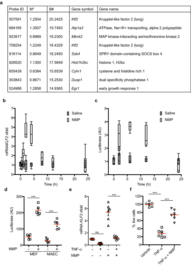
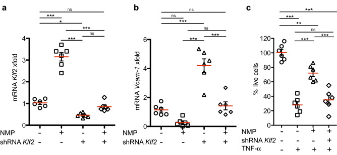
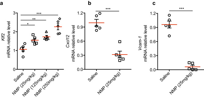
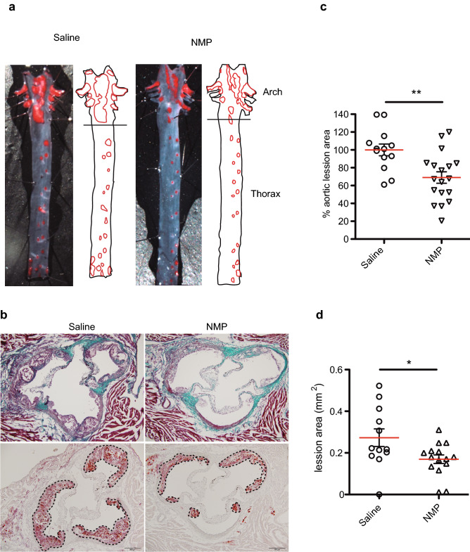
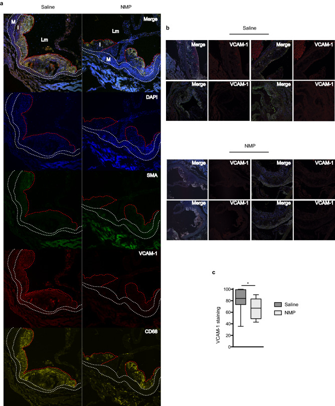
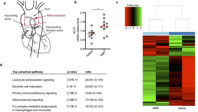
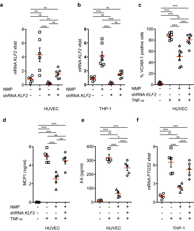
N-methyl-2-pyrrolidone (NMP) is a versatile water-miscible polar aprotic solvent. It is used as a drug solubilizer and penetration enhancer in human and animal, yet its bioactivity properties remain elusive. Here, we report that NMP is a bioactive anti-inflammatory compound well tolerated in vivo, that shows efficacy in reducing disease in a mouse model of atherosclerosis. Mechanistically, NMP increases the expression of the transcription factor Kruppel-like factor 2 (KLF2). Monocytes and endothelial cells treated with NMP express increased levels of KLF2, produce less pro-inflammatory cytokines and adhesion molecules. We found that NMP attenuates monocyte adhesion to endothelial cells inflamed with tumor necrosis factor alpha (TNF-α) by reducing expression of adhesion molecules. We further show using KLF2 shRNA that the inhibitory effect of NMP on endothelial inflammation and subsequent monocyte adhesion is KLF2 dependent. Enhancing KLF2 expression and activity improves endothelial function, controls multiple genes critical for inflammation, and prevents atherosclerosis. Our findings demonstrate a consistent effect of NMP upon KLF2 activation and inflammation, biological processes central to atherogenesis. Our data suggest that inclusion of bioactive solvent NMP in pharmaceutical compositions to treat inflammatory disorders might be beneficial and safe, in particular to treat diseases of the vascular system, such as atherosclerosis.


Conflict of interest statement

The authors declare no competing interests.

Figures

Figure 1
Figure 1
Bioactive NMP is a Klf2 transcriptional activator. (a) Microarray analysis showing top 10 genes up regulated in C2C12 cells by addition of 5 mM NMP for 8 h. (b) Real-time PCR analysis of Klf2 mRNA in mouse C2C12 cells at different time points after NMP administration. Klf2 mRNA amounts are normalized to Gapdh mRNA and are presented relative to the level observed in untreated cells. Data are presented as mean ± SEM; n = 6. (c) C2C12 cells transfected with a luciferase Klf2-promoter reporter analyzed at different time points w/wo NMP (1 mM). The luciferase activity was measured and normalized to Renilla activity, and represented in arbitrary units (AU) from two independent assays (n = 6). (d) MEF and MAEC cells transfected as in (c) were treated with or without NMP. Luciferase assays were performed 24 h after transfection. Data are presented as mean ± SEM; n = 5 *** p < 0.001 (one-way ANOVA with the Bonferroni comparison post-test). (e) TNF-α mediated (10 ng/ml) inhibition of Klf2 mRNA is suppressed by NMP (1 mM). Data are presented as mean ± SEM; n = 6, ns p > 0.05, *** p < 0.001, one-way ANOVA. (f) NMP pretreatment for 6 h protects against cell death induced in C2C12 cells activated with TNF- α for 24 h (n = 6; ***p < 0.001, one-way ANOVA with the Bonferroni comparison post-test).
Figure 2
Figure 2
NMP ameliorates TNF-α-induced-apoptosis through the Klf2 pathway. (a) Exponentially growing non-synchronized cells were transduced with lentiviral particles encoding for shRNA against KLF2 mRNA (“shRNA”) or for a non-specific shRNA as control; 72 h after infection cells stimulated with TNF- α for 6 h were exposed to NMP (1 mM), RNA was purified and Klf2 (a) and Vcam-1 (b) mRNA levels were analyzed by QPCR, and (c) cell viability was measured by flow cytometry. The results in C2C12 cell line show at least two independent biological replicates (n = 6). Error bars represent the standard error of the means (SEM). Significant differences are represented by asterisks: *p < 0.05, **p < 0.01 and ***p < 0.001, or non-significant (ns), by one-way ANOVA with Bonferroni’s multiple comparison test.
Figure 3
Figure 3
NMP is bioactive in vivo. (a) Real-time PCR analysis in liver samples of mRNA expression of (a) Klf2 mRNA in mice treated with 25, 125 and 250 mg/kg of NMP. ***p < 0.001, **p < 0.005,*p < 0.01 by one-way ANOVA with Bonferroni’s multiple comparison test (n = 5 mice). Each data point denotes a liver sample from an individual mouse. (b,c) endogenous mouse Cxcl12 and Vcam-1 mRNA in treated mice with 25 mg/kg mouse. mRNA amounts are normalized to Gapdh mRNA relative to the level of liver samples in non-treated animals. (n = 5; ***p < 0.001, unpaired Student’s t test).
Figure 4
Figure 4
NMP attenuates atherosclerosis development in ApoE − / − mice. (a) Representative en face Oil Red O staining of aortas from treated and untreated NMP mice maintained on HFD for 12 weeks. (b) Representative Masson’s trichrome (upper panel) and Oil Red O (lower panel) staining in aortic root sections from HFD-fed NMP treated and untreated mice. Scale bar, 200 μm. (c) Quantitative analysis of atherosclerotic lesion size in Oil Red O-stained in aortic arch region from (a). (d) Quantitative analysis of atherosclerotic lesion size in Oil Red O-stained aortic sections from (c). (n = 12–18; *p < 0.01, **p < 0.005, unpaired Student’s t test).
Figure 5
Figure 5
NMP treatment reduces macrophages infiltration and inflammation. (a) Representative immunostaining of macrophages, smooth muscle cells (SMC) and activated endothelium in aortic sinus lesions of NMP treated and untreated C57BL/6 J mice fed with HFD for 12 weeks. Lesions were stained for biomarkers of macrophages (CD68; yellow), SMCs (α-actinin; green) and stressed endothelium (Vcam-1; red); nuclei were stained with DAPI (blue). Merged images are also shown. Lm, aortic lumen, M, tunica media, and I, tunica intima. White dashed lines demarcate the elastic lamina. Bars, 200 μm. (b) Vcam-1 and merge staining as in (a) from different animals. (c) Quantitative analysis of relative Vcam-1 immunostaining relative to the aortic sections size from (b).
Figure 6
Figure 6
Gene expression signature of NMP treatment in the atherosclerotic aorta. (a) The diagram shows the aortic section plane (red line) and the aortic tissue sampled for RNA-seq studies. (b) Real-time PCR analysis of Klf2 mRNA in aortic arches after NMP or saline administration. Klf2 mRNA amounts are normalized to Gapdh mRNA and are presented relative to the level observed in untreated aortas. (c) Heat map of top genes differentially expressed in aortic arches between NMP and saline treated mice. Scaled expression values are color-coded according to the legend on the left. The dendrogram depicts hierarchical clustering based on the top differentially expressed genes. The top bar indicates the treatment: light blue, NMP; blue, saline control. (d) Top canonical pathways that were significantly deregulated by NMP treatment, as identified by Ingenuity Pathway Analysis. The significance of association between altered genes and the canonical pathway was assessed using a right-tailed Fisher's exact test to calculate a p value determining the probability that the association is explained by chance alone. Ratios defining the proportion of deregulating genes from a pathway related to the total number of molecules that make up that particular pathway are also indicated.
Figure 7
Figure 7
NMP inhibits TNF-α-dependent vascular inflammation by KLF2. (a) Real-time PCR analysis of KLF2 mRNA in human HUVEC and (b) THP-1 cells after 24 h of NMP administration. KLF2 mRNA amounts are normalized to GADPH mRNA and are presented relative to the level observed in untreated cells. (c) Percentage of VCAM-1 positive HUVEC cells. NMP reduces cytokine release in HUVEC cells after TNF-α stimulation. Supernatants from HUVEC cells were harvested after 24 h of treatment, and the concentration of MCP-1 (c) and IL-6 (d) measured by ELISA, TNF-α treated with NMP compared with TNF- α untreated cells. (f) Real-time PCR analysis of PTGS2 mRNA in human THP-1 cells. All data is shown as mean ± SEM (n = 4–7). *p < 0.01, **p < 0.005, ***p < 0.001 by one-way ANOVA with Bonferroni’s multiple comparison test.
Figure 8
Figure 8
NMP attenuates THP‐1 monocyte adhesion to HUVECs. (a) Representative frames (from Supplemental Video 1) showing THP-1 cells adherence to a monolayer of TNF-α pre-activated HUVECs after treatment with NMP or saline. Numbers indicate elapsed time in minutes. (b) Quantitative analysis of the percentage of remaining attached THP-1 cells to the HUVEC monolayer by comparison of the percentage of attached cells at the beginning of the flow (time = 0 min). (c) Representative immunostaining of attached PBMC to a TNF-α activated HUVEC cells monolaye after 5 min of flow (5 dyn/cm2). PBMCs were stained with CD11b in green, and stressed endothelium with VCAM-1 (red), nuclei were stained with DAPI (blue). Merged images are also shown. (d) Quantitative analysis of VCAM-1/DAPI positive staining area from (c). Quantitative analysis of CD11b positive stained cells per field from (C). (n = 5; *p < 0.01, ***p < 0.001, unpaired Student’s t test).

References

    1. Domanski M, Lloyd-Jones D, Fuster V, Grundy S. Can we dramatically reduce the incidence of coronary heart disease? Nat. Rev. Cardiol. 2011;8:721–725. doi: 10.1038/nrcardio.2011.158. - DOI - PubMed
    1. Libby P, Ridker PM, Hansson GK. Progress and challenges in translating the biology of atherosclerosis. Nature. 2011;473:317–325. doi: 10.1038/nature10146. - DOI - PubMed
    1. Back M, Yurdagul A, Jr, Tabas I, Oorni K, Kovanen PT. Inflammation and its resolution in atherosclerosis: mediators and therapeutic opportunities. Nat. Rev. Cardiol. 2019;16:389–406. doi: 10.1038/s41569-019-0169-2. - DOI - PMC - PubMed
    1. Ridker PM, Luscher TF. Anti-inflammatory therapies for cardiovascular disease. Eur. Heart J. 2014;35:1782–1791. doi: 10.1093/eurheartj/ehu203. - DOI - PMC - PubMed
    1. Martinez-Hervas S, Gonzalez-Navarro H. Anti-inflammatory therapies for cardiovascular disease: signaling pathways and mechanisms. Rev. Esp. Cardiol. (Engl. Ed.) 2019;72:767–773. doi: 10.1016/j.rec.2019.03.007. - DOI - PubMed

Publication types

MeSH terms